
Hanoi Ms. Thuy, 78 years old, had colon cancer surgery three years ago. This time, during a routine health check-up at Tam Anh General Hospital, a colon tumor was discovered, which needed to be removed endoscopically to be completely removed.
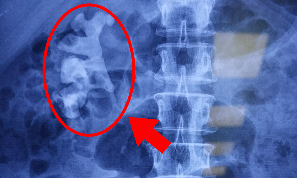
The dye endoscopy results showed many polyps, in the transverse colon of the right hepatic flexure there was a large lesion spreading to both sides (5.5x4 cm). The dye endoscopy method and narrow band imaging (NBI) technique helped the doctor observe more clearly the vascular pattern and pit tissue (two signs of malignancy). Thanks to that, the doctor can evaluate polyps and lesions with a high risk of cancer invading the submucosa.
On March 3, Dr. Dao Tran Tien, Deputy Head of the Gastroenterology Department, Tam Anh General Hospital, Hanoi, said that this is a high-grade dysplastic tumor, in the pre-cancerous stage. Previously, for pre-cancerous tumors or early-stage cancer, doctors often performed surgery to remove a segment of the colon. As for the elderly patient Thuy, who had previously had surgery to remove half of her colon, a second surgery would easily lead to complications, a risk of losing colon function due to complete removal, reducing her quality of life.
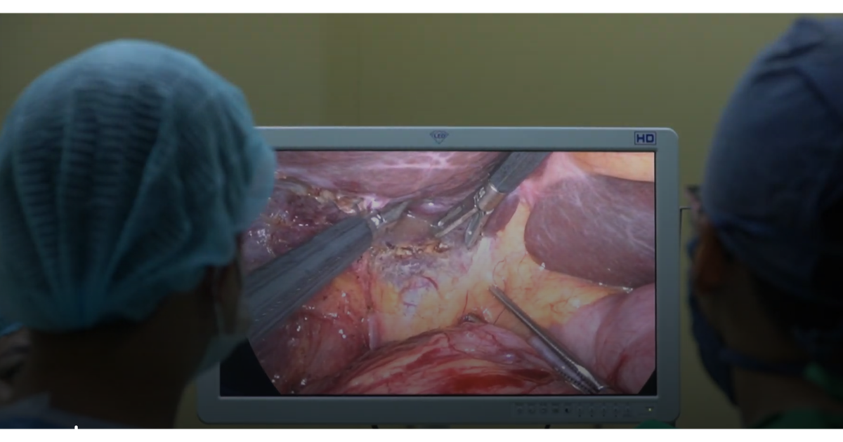
After a multidisciplinary consultation, the doctor chose the ESD (endoscopic mucosal dissection) method. The team inserted the endoscope from the anus through the colon to the lesion, used specialized instruments to separate the mucosa below, and removed the dysplastic lesion.
According to Dr. Tien, the patient had a history of sigmoid colon cancer, had undergone surgery and chemotherapy, so there were changes in anatomy and high adhesions, so the team needed to handle it skillfully and thoroughly cut out the lesion at the bottom of the mucosa. In addition, the elderly patient had underlying hypertension and a thin colon, so the doctor had to be careful in every operation.
The patient had the colon tumor completely removed by mucosal resection, and 9 polyps scattered throughout the colon were removed during endoscopy. The doctor clamped and cauterized the bleeding points, closed the wound, and minimized the rate of local complications.
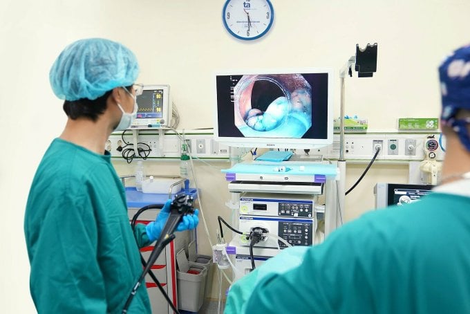
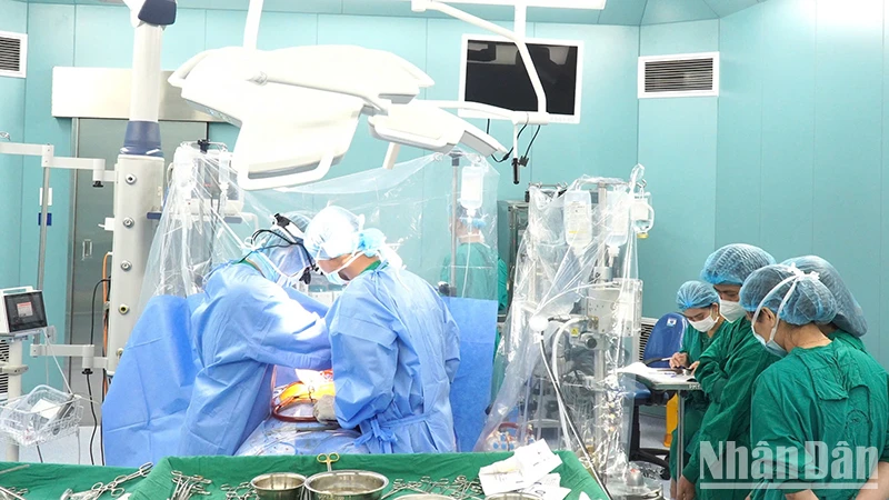
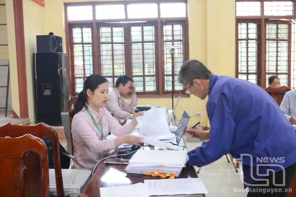
Doctor Tien (left) performs an endoscopy to remove a lesion in Ms. Thuy's colon. Photo: Provided by the hospital
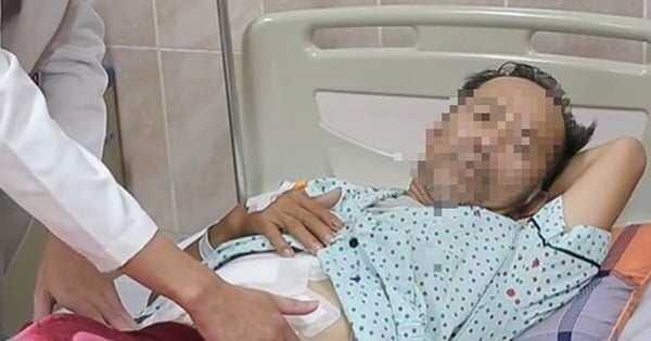
One day after surgery, Ms. Thuy practiced eating porridge again, her health was stable, no abdominal pain or bloating, and she was discharged from the hospital after three days.
Dr. Tien noted that patients who have undergone colon mucosal resection should eat soft, easily digestible foods such as porridge, soup, and pureed fiber. Limit strenuous exercise related to the area near the intervention site.
Colon cancer is quite common, with a high incidence in people aged 40-50. More than half of colon cancer cases occur in the rectum and sigmoid colon. Colon tumors have no symptoms and can easily turn into cancer. When a tumor is detected, the patient should have a regular check-up every 6 months to promptly detect and treat any progressive lesions, if any.
Emerald
* Patient's name has been changed
| Readers ask questions about digestive diseases here for doctors to answer |
Source link


Comment (0)