
Capsular contracture only occurs in women after breast implants are placed, fibrous tissue will begin to form as a shell around the implant called "pocket". Simply put, this will be a protective shell that protects the implant independent of the body's tissues after placement.
These tissues are usually very soft, but some people after surgery may experience excessive proliferation, reduced organization of fibrous tissue around the implant, forming a fibrous capsule, hard scar tissue, and causing capsular contracture after breast augmentation, which can cause pain and deform the shape of the breast.
Specifically, there is a defect or bulge in a certain position on the breast, the breast protrusion is high or low, the breast base is high or low, the breast cleavage becomes wide and uneven, and even severe cases cause distortion of the shape of the entire breast.
Therefore, capsular contracture is a concern for anyone preparing for and after breast augmentation surgery. Below is the sharing of Master, Doctor Ho Cao Vu, currently working at the Department of Surgery, Cho Ray Hospital, about the causes, signs, and surgical methods of removing the fibrous capsule to reconstruct the chest cavity using an ultrasonic scalpel.
Remove breast implants and peel grade 4 fibrous capsule.
Causes of capsular contracture
The cause of capsular contracture does not come much from the breast implant but mostly from the surgical technique.
The doctor designed the pocket to be narrow compared to the size of the breast implant: In cases where the pocket is narrow, there are two reasons.
First, the customer chose a breast implant that was too large for the body's anatomical structure, but the doctor did not advise the customer on the appropriate size of the implant. The surgeon designed the wrong size of the implant cavity, which was too narrow or did not match the customer's chest structure, causing pressure on the implant over time, shrinking the implant, leading to fibrosis.
Causing damage during cavity creation: In all pathological and cosmetic surgeries, the surgery is too rough, causing damage, leading to the patient having a lot of pain after surgery, a lot of blood and secretion, slow healing and late complications such as fibrosis.
Signs of fibrosis
Grade 1: The breast is still soft and looks normal, the capsule is slightly firm when touched in the supine position.
Level 2: The breast looks normal, no swelling or pain, no deformity, but feels harder than normal, especially in the supine position.
Level 3: The breast is hard and deformed due to contraction, it may be round or the breast implant is pulled upwards and downwards causing deformation, there is a continuous dull pain in the chest area, and breast deformation.
Level 4: The breast is completely distorted, misaligned, completely asymmetrical, the fibrous capsule is very hard, constricted and causes a lot of pain, constant discomfort in the chest area.
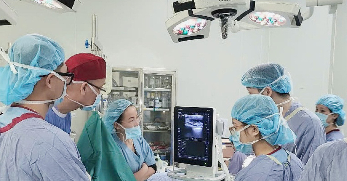
Use ultrasound to remove breast implants and peel off fibrous capsule.
Why is an MRI scan needed before capsulotomy?
In-depth breast MRI before removing breast implants and peeling off the fibrous capsule helps check for breast pathology, fibrous capsule, pocket, diseases related to breast implants, tumors, etc. (Recommended: regular MRI, ultrasound, X-ray are not effective in cases of breast implants and breast implant rupture).
The doctor will then assess the condition of the fibrous capsule, any abnormalities inside and outside the pocket and determine the best surgical method for the patient. In cases of double fibrous capsules and difficult separation, the surgery can last up to 4 hours - the doctor will reshape the cavity to place the new pocket.
For major breast implant manufacturers in the US, a detailed breast MRI is one of the necessary factors to help customers get new implants, if still under warranty.
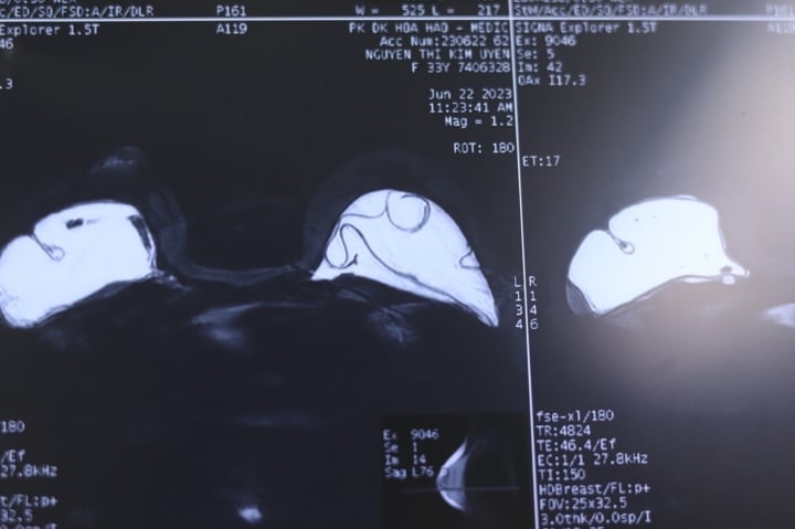
MRI results show breast implant rupture and capsular contracture.
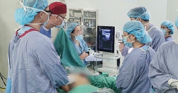
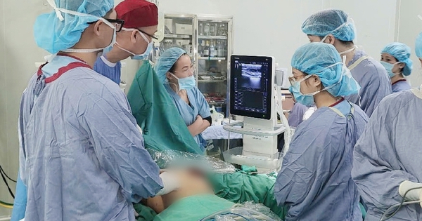
Breast implant removal and capsule peeling procedure using ultrasonic knife
Step 1: In-depth MRI of the breast to check for breast disease, breast implants, pockets and diseases related to breast implants.
Step 2: Screen for possible cancer risk and lesions related to the bag.
Step 3: Clinical examination to plan breast implant removal surgery, handle associated complications such as fibrous capsule, ruptured implant, slipped implant, leaking implant, etc. And determine whether to replace with a new implant?
Step 4: Health check-up, women need to perform tests before breast implant removal surgery at a specialized general hospital. For normal breast implant removal cases, it only takes 30-45 minutes, but for breast implant removal cases that require difficult fibrous capsule peeling, re-creating the implant cavity, or implant rupture, the anesthesia time will be longer.
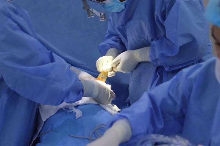
Step 5: Breast implant removal surgery, the doctor makes a 3cm - 3.5cm incision at the areola or chest base (in cases where there are no abnormalities) and uses a Harmonic or Innolcon, Enseal, Ligasure ultrasonic knife to cut the tissue inside to remove the old breast implant. Check the implant brand, size, implant size and projection.
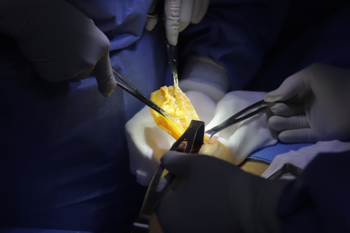
Remove the textured bag during breast implant removal and capsulectomy.
Step 6: Clean the pocket cavity. In cases of ruptured breast implants or abnormal fluid, the team must clean the pocket cavity, culture the fluid, and perform an antibiotic test - when the fluid is cloudy.
Step 7: Use an ultrasonic knife to peel off the fibrous capsule and take fibrous tissue for pathological examination to determine whether the fibrous tissue is benign or malignant. Prepare a cold biopsy if malignancy is suspected.
Step 8: Reshape the pocket (replace a new pocket if indicated) and correct the pocket to improve the condition of the gap, pocket slippage, or the pocket being too wide or too narrow.
Step 9: Wear a post-operative shaping garment. In cases where the bag is removed and replaced, the patient usually does not drain on the same day. In cases where the bag is removed with abnormalities such as peeling of the fibrous capsule or severe internal damage, the patient will need to drain and stay in the hospital for 1 night before being discharged.
Advantages of using ultrasonic scalpel to peel off fibrous capsule
Dr. Ho Cao Vu has more than 10 years of experience using ultrasonic knives in pathological and cosmetic surgery. He shared: removing breast implants, peeling off the fibrous capsule and replacing a new implant when using an ultrasonic knife is similar to when performing a new breast augmentation with advantages such as no bleeding, no pain, quick healing, no long-term scarring, no need to rest, no painkillers or antibiotics and can go home the same day without having to stay in the hospital.
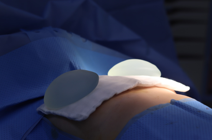
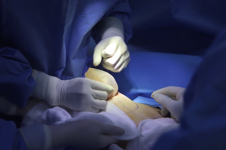
Image of breast implants after removal from the body.
Below is the case of a customer 1m55 tall, 50kg in weight who had breast augmentation 6 years ago at a famous cosmetic hospital in Ho Chi Minh City. Currently, feeling that her breasts are abnormally drooping and hard when lying down, she came to the cosmetic hospital for a re-examination, hoping to remove the breast implants.
Receiving unreasonable advice, she decided to have an in-depth breast MRI, with the results of ruptured breast implants and fibrous capsule. She went to Dr. Ho Cao Vu to decide to remove the ruptured breast implants, peel off the fibrous capsule and replace them with new breast implants.
During the surgery, the surgeon dissected layer by layer into the cavity where the two bags were placed. The cavity on the left side of the bag had ruptured, the gel in the cavity had turned yellow, the bag shell was aging and very fragile, the doctor took out the ruptured bag, pumped and cleaned it, and checked that the bag shell was covered with a fibrous capsule with rough spots on the surface, and a double fibrous capsule on the back.
The right cavity of the ruptured sac had yellow gel, the sac was aged and crumbly, the doctor took out the entire sac and gel, washed the cavity, there was a fibrous capsule in the lower area outside the cavity and the back had many papillomas, peeled off part of the fibrous capsule on both sides, recreated the cavity, cut the fibrous capsule area with papillomas on the back right side and sent it for cytological testing.
Bao Anh
Source

![[Photo] Prime Minister receives a number of businesses investing in Ba Ria-Vung Tau province](https://vstatic.vietnam.vn/vietnam/resource/IMAGE/2025/3/20/8e3ffa0322b24c07950a173380f0d1ba)
![[Photo] President Luong Cuong receives former Vietnam-Japan Special Ambassador Sugi Ryotaro](https://vstatic.vietnam.vn/vietnam/resource/IMAGE/2025/3/20/db2d8cac29b64f5d8d2d0931c1e65ee9)


![[Photo] President Luong Cuong receives Ambassador of the Dominican Republic Jaime Francisco Rodriguez](https://vstatic.vietnam.vn/vietnam/resource/IMAGE/2025/3/20/12c7d14ff988439eaa905c56303b4683)


Comment (0)