
Valvular heart disease is rapidly increasing and is a common cause of cardiovascular death.
For patients with heart valve regurgitation, timely treatment helps prevent dangerous complications such as atrial fibrillation, heart failure, blood clots, endocarditis, abnormal heart rhythms, stroke and death.
 |
| Illustration |
According to doctors, the heart has 4 valves including: 2-leaf valve, 3-leaf valve, aortic valve and pulmonary valve. Heart valves help blood flow in one direction, patients with heart valve regurgitation, blood flows in the opposite direction.
The mitral valve functions to direct blood from the left atrium into the left ventricle and prevent backflow from the left ventricle to the left atrium. If the mitral valve is leaky, blood will flow back from the left ventricle back to the left atrium during systole. A malfunctioning heart valve that does not close and open properly will affect the heart's ability to pump blood to the body. Thus, the heart valve plays a very important role.
Common heart valve diseases include: Valve stenosis or valve regurgitation. The older the patient, the higher the risk of heart valve regurgitation or valve disease.
In aortic disease, the older you get, the more the aorta dilates, causing valve regurgitation. Possible causes of valve regurgitation include: degenerative valve, infective endocarditis, genetics, etc.
In case the patient has no symptoms, when going for a routine health check-up, an echocardiogram will be ordered. If the results show mitral valve or aortic regurgitation ¼ or 2/4, medical treatment will be indicated.
However, before that, the doctor will find out the cause of the valve regurgitation. In case of drug treatment, the patient will still be monitored annually, every 6 months, or examined when tired, short of breath, or heart palpitations when exerting.
Heart valve regurgitation can be detected even in patients who only go for a general health check-up, they have no symptoms and ultrasound accidentally detects heart valve regurgitation.
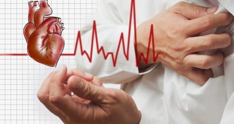
If one of the four heart valves is severely leaky, the patient may experience symptoms such as: reduced ability to exert, fatigue, chest pain, anxiety, rapid heartbeat, dizziness, fainting... These are common signs of heart valve disease.
If a patient with severe heart valve regurgitation is not detected promptly, it can lead to complications including:
Heart failure, reduced left ventricular ejection fraction as well as right ventricular failure. Dangerous arrhythmias, reduced quality of life, increased mortality.
Risk of infection from oral cavity, bacteria can enter the damaged heart cavity through the blood, leading to complications of infective endocarditis, which can lead to stroke or embolism in all blood capillaries in the organ. The patient can go into septic shock and die.
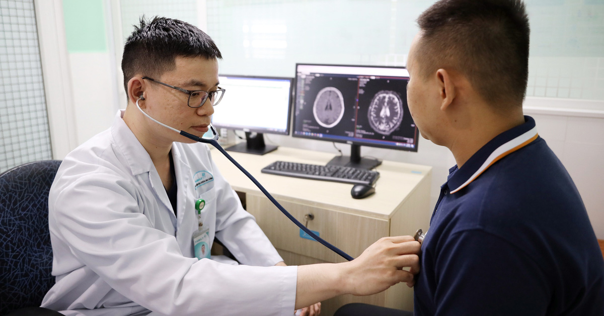
According to Master Tran Thuc Khang, Cardiovascular Center, Tam Anh General Hospital, Ho Chi Minh City, heart valve surgery is basically still open heart surgery.
This means that during surgery, the heart stops beating and the patient's circulation is nourished by a heart-lung machine outside the body. In current open heart surgery, to treat heart valve disease, the surgeon can repair or replace one or more diseased heart valves through the skin.
Minimally invasive techniques, that is, surgery through a small incision in the right chest, combined with a minimally invasive television support system, are increasingly interested and more widely used, especially in mitral valve diseases.
However, not all heart valve diseases can be treated with invasive techniques. The choice of when to perform open surgery or invasive surgery depends on many factors.
For example, surgery on one valve or multiple valves, whether mitral valve surgery is accompanied by coronary artery disease or not, whether the patient's aorta is dilated or not, whether the patient's chest has been previously secured or not, whether the patient is obese or not, whether the heart failure is too severe or not, whether the aorta, iliac arteries and the arteries of both lower limbs are pathological or not.
In invasive techniques, the patient needs to be put on extracorporeal circulation through the cephalic aorta. Thus, before choosing a surgical method, the surgeon must examine, evaluate the patient and discuss directly with the patient the advantages of that method.
For minimally invasive techniques, there are many advantages and safety similar to open surgery. Some outstanding advantages are: Less pain, shorter surgical scars, patients do not need to have surgery along the middle of the sternum, so the recovery time is faster. At the same time, complications related to the surgical line, especially bleeding and infection, will be less. Thanks to that, the patient's hospital stay will be shorter and the cost will be lower.
This is an ultrasound-guided anesthesia technique. The anesthesiologist will insert a catheter (a small tube) into the space between the erector spinae muscles, which are the muscles on either side of the patient's spine. The catheter has a syringe system and an automatic pump.
In the heart pump, the doctor will prepare a certain dose of medicine according to a protocol and the anesthetic will be released within 48 to 72 hours after surgery. The anesthetic penetrates the surface of the erector spinae muscle, the nerve roots in the erector spinae muscle will block the central nervous signals passing through the spinal cord scar horns. From there, it helps the patient reduce pain.
According to Dr. Khang, this method offers the advantage of very good post-operative pain relief. Previously, cardiothoracic surgery often used post-operative pain relief with intravenous morphine preparations.
If the dose is high, morphine will cause respiratory depression, complications of urinary retention, vomiting, and even some patients with hyperpneumonia will develop dependence and addiction to morphine. The technique of erector spinae plane block helps reduce the dose of morphine used after surgery, thereby reducing complications related to morphine.
According to Dr. Nguyen Duc Hung, Deputy Head of Cardiology Department, Tam Anh General Hospital, Hanoi, not all lesions are suitable for percutaneous surgery.
Therefore, before proceeding with transcutaneous valve repair or replacement, the patient needs to be examined, tested and thoroughly non-invasively to ensure anatomy. Because if the valve defect is suitable, then transcutaneous valve repair can be performed.
For other valve regurgitation such as pulmonary valve, if pulmonary regurgitation occurs after congenital open heart surgery or natural regurgitation, percutaneous pulmonary valve replacement can be performed.
Or tricuspid valve regurgitation can be repaired or replaced through the skin. The difference between percutaneous valve replacement and other techniques is specifically in the access route of the technique.
During percutaneous valve replacement, we will open a blood vessel in the thigh. From that access point, we will introduce instruments to access specific heart chambers such as the mitral valve, pulmonary valve, and tricuspid valve.
Because it is minimally invasive, this method helps patients recover quickly, reduces bleeding, and reduces infection. However, it is necessary to carefully evaluate whether this solution is suitable for the patient or not, before making a decision to discuss and advise the patient.
Source: https://baodautu.vn/tang-nhanh-benh-ly-van-tim-d225691.html


![[Photo] Solemn Hung King's Death Anniversary in France](https://vstatic.vietnam.vn/vietnam/resource/IMAGE/2025/4/6/786a6458bc274de5abe24c2ea3587979)
![[Photo] Vietnamese rescue team shares the loss with people in Myanmar earthquake area](https://vstatic.vietnam.vn/vietnam/resource/IMAGE/2025/4/6/ae4b9ffa12e14861b77db38293ba1c1d)


Comment (0)