
Medical news on January 9: Risk of acute pancreatitis and kidney stones at the end of the year
At the end of the year, when festivals, parties, and partner meetings take place continuously, many people face serious health problems, including acute pancreatitis.
Acute pancreatitis from alcohol and irregular living habits
Acute pancreatitis is an acute inflammatory lesion of the pancreas, leading to systemic inflammation, causing disorders of many organs such as the heart, lungs, liver, kidneys, and severe cases can lead to many complications such as respiratory failure, blood clotting disorders, septic shock, etc.
 |
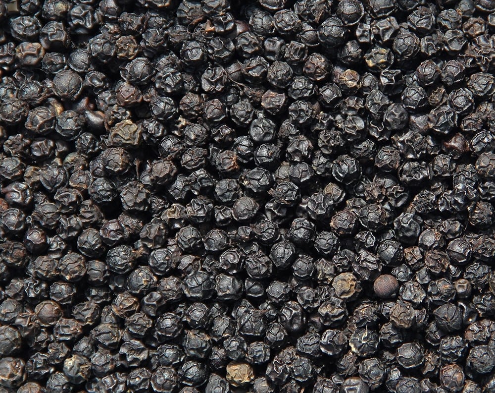
| Acute pancreatitis is a condition that people often encounter when abusing alcohol. |
Acute pancreatitis allows activated enzymes and toxins such as cytokines to spill out of the pancreas, into the abdominal cavity causing peritonitis, septic shock and spreading to other organs causing multiple organ failure. Toxins can be absorbed from the abdomen into the lymphatic vessels, then into the blood causing hypotension, sepsis, and damage to organs outside the abdominal cavity.
Worldwide, alcohol is a common cause of pancreatitis. Acute alcoholic pancreatitis occurs mainly in men, especially middle-aged men (40 years and older) with a history of alcohol abuse (drinking heavily and regularly).
Initial symptoms include severe epigastric pain that may radiate to the back, accompanied by bloating and vomiting. In mild cases, the pain may be mild, dull, and last for 2-3 days.
In severe cases, the disease progression is often acute, with symptoms of severe pain, stabbing sensation, abdominal distension, fever... and in severe cases, the patient's risk of death increases by about 10-30%.
Less commonly, pancreatitis develops silently and lasts for a long time without any symptoms such as abdominal pain or vomiting. It is usually only diagnosed when it affects pancreatic function such as diabetes, or digestive disorders, fatty stools, or pancreatic pseudocysts.
Pancreatitis can manifest in acute or chronic form with varying degrees of severity. To diagnose acute pancreatitis, doctors often rely on the patient's clinical symptoms such as typical abdominal pain, bloating, vomiting combined with increased blood pancreatic enzyme tests (increased amylase, lipase) or images of pancreatitis on ultrasound or abdominal CT scan.
In addition to a definitive diagnosis of pancreatitis, patients also need to have blood tests to determine the severity of pancreatitis as well as the cause of pancreatitis in each patient. Recurrent episodes of acute pancreatitis, such as Tuyen's case, require detailed examination to determine the cause.
Recurrent acute pancreatitis can cause persistent inflammation, which over time leads to changes in the pancreatic parenchyma such as pancreatic parenchyma atrophy, fibrosis, parenchymal calcification, or pancreatic stones, becoming chronic pancreatitis.
Pancreatitis is a serious disease that can be fatal if not detected and treated promptly, or if not monitored and treated thoroughly, can leave many complications. Complications of pancreatitis not only affect life but also greatly affect the quality of life.
According to Dr. Dao Tran Tien, Deputy Head of the Department of Gastroenterology, Tam Anh General Hospital, Hanoi, acute complications of acute pancreatitis such as necrotizing pancreatitis, hypovolemic shock, or organ failure such as kidney failure, respiratory failure, etc. can occur in severe cases of acute pancreatitis, increasing the risk of death in patients from 2-10% and severe cases of acute pancreatitis after treatment need to be monitored and treated to prevent progression to pancreatic pseudocysts and pancreatic abscesses.
Cases of pancreatitis that recur, progress over a long period of time, or are not treated thoroughly can lead to complications such as chronic pancreatic insufficiency leading to reduced production of pancreatic digestive enzymes, leading to exhaustion, malnutrition, or impaired endocrine pancreatic function leading to complications of diabetes due to the pancreas.
Acute pancreatitis is best prevented by avoiding factors that are the cause or risk of pancreatitis such as limiting alcohol (causing direct damage or infection affecting pancreatic function), preventing gallstones (bile duct stones, gallbladder stones), diabetes (people with diabetes have a 30% higher risk of acute pancreatitis);
Limit the use of drugs that can cause pancreatitis (nonsteroidal anti-inflammatory drugs, or steroids), control dyslipidemia (increased triglyceride levels in obese people) or treat other endocrine diseases such as hyperparathyroidism or high blood calcium or screen people with a family history of pancreatitis...
In particular, people with a history of pancreatitis should limit alcohol (cut down or quit drinking); avoid eating too much protein and fat in one meal (especially during Tet), follow a balanced diet (drink enough water, enough protein, eat lots of fruits and vegetables), exercise regularly, maintain a healthy weight (losing weight if overweight can help reduce the risk, limit fat); avoid smoking and should have regular check-ups so that doctors can monitor and advise on health status.
53-year-old patient with large coral stones causing kidney failure
Ms. NTTV, 53 years old, living in Khanh Hoa, had been suffering from back and hip pain for two months without knowing the cause. The pain often appeared when she bent over or did strenuous work, making her quickly feel tired and had to lie on her right side to relieve the pain. In addition, she also discovered that her urine was cloudy and had an unpleasant odor. Worried, she decided to go to the hospital for a health check.
At the hospital, she was assigned by Dr. Nguyen Truong Hoan, Department of Urology, Center for Urology - Nephrology - Andrology, to have a computed tomography (CT-scan) scan to examine her lower back area.
The results showed that her left kidney was hydronephrotic and had a large coral-shaped stone, consisting of 4 branches spreading into the renal calyces. The total size of the stone was up to 5-6 cm, occupying about ⅓ of the volume of the left kidney. In addition, she also had a urinary tract infection.
This type of coral stone not only causes urinary tract obstruction but also causes hydronephrosis, leading to kidney failure if not treated promptly. This is a case of infected coral kidney stones, a very dangerous type of urinary stone.
With infected coral kidney stones, antibiotics are needed to control the infection before surgery. Ms. V. was treated with antibiotics for a week and had a urine culture done to ensure that the infection was completely controlled. If the infection is not treated before the stone is crushed, bacteria from the stone can enter the bloodstream, which is life-threatening.
After the urine culture results were negative and the infection was stable, Ms. V. was scheduled to undergo mini PCNL.
This is the optimal method for treating large coral stones, with outstanding advantages such as less bleeding, less surgical site infection and less post-operative pain, helping patients recover quickly.
During the surgery, with the help of ultrasound and the C-Arm system to accurately locate the stone, the doctors created a small passage of less than 1 cm from the outside of the skin on the left flank to the inside of the renal pelvis. Then, the stone was approached and crushed into small fragments by high-power laser energy, then sucked out.
After about 180 minutes, the entire coral stone mass was removed from Ms. V's left kidney. One day after the surgery, Ms. V. recovered quickly, no longer felt pain, and could eat and move normally. After a week of follow-up examination, the ultrasound results showed that her left kidney was completely free of stones.
Coral stones account for only about 10-15% of urinary stones, but are the most dangerous type of stones. Coral stones often develop in urinary tract infections and easily cause hydronephrosis, urinary tract obstruction, and impaired kidney function. If not treated promptly, coral stones can lead to kidney infections, pyelonephritis, kidney failure, and even life-threatening blood infections.
Coral stones often develop silently, with few symptoms or only signs such as lower back pain, cloudy urine, fatigue, etc. Therefore, Dr. Hoan recommends that people with a history of kidney stones, especially coral stones, should proactively have regular health check-ups every 6-12 months to detect kidney stones early when they are small and can be treated with less invasive methods, such as medication or extracorporeal lithotripsy.
With the percutaneous endoscopic lithotripsy (mini PCNL) treatment, Ms. V. was able to safely and effectively treat her coral kidney stones. This is a typical example showing that early detection and treatment of kidney stones can help patients avoid dangerous complications and recover quickly.
Gene mutation causes postpartum heart failure in mothers
Ms. Nhi, 41 years old, had to go through a difficult journey when she suddenly gained more than 10kg, her legs were swollen and she had difficulty breathing, even when doing normal activities. After examination, she was diagnosed with severe heart failure due to peripartum cardiomyopathy.
Ten years ago, after giving birth to her second daughter, Nhi began to experience symptoms such as fatigue, shortness of breath, and swelling of the legs. Initially, she was diagnosed with unexplained heart failure and treated as directed by her doctor. After a while, she felt better, went about her daily activities and worked normally, but stopped taking her medication on her own and skipped follow-up visits.
By early 2024, Ms. Nhi's symptoms had strongly relapsed with symptoms of difficulty breathing at night, shortness of breath when walking and doing activities, along with rapid weight gain (12kg in less than 1 month). She decided to go to a large hospital for a health check.
MSc. Do Thi Hoai Tho, Heart Failure Clinic, Cardiovascular Center, said that Ms. Nhi was hospitalized with swelling of the face and legs, fatigue and severe shortness of breath.
Echocardiography showed a left ventricular ejection fraction (LVEF) of only 13% (normal > 50%), indicating severe heart failure. Coronary angiography showed no signs of obstruction, but cardiac MRI showed signs of dilated cardiomyopathy.
Genetic testing revealed that Nhi carried a mutation in the TTN gene. This mutation is thought to be responsible for approximately 20% of cases of dilated cardiomyopathy in families. Women with the TTN gene mutation who become pregnant and give birth are at increased risk of developing peripartum cardiomyopathy, a form of dilated cardiomyopathy.
Peripartum cardiomyopathy is a rare condition that occurs during the last months of pregnancy and up to 5 months after birth. It causes a weakening of the heart's contractile function, leading to heart failure. It is particularly common in women over 30 and can be caused by a number of factors, including hormonal changes during pregnancy, viral myocarditis, and genetic mutations.
Upon admission, Ms. Nhi had to use oxygen and stay in bed due to severe heart failure. After examining and determining the cause, the doctor prescribed treatment with diuretics combined with the basic medicine for heart failure. After more than a week of treatment, Ms. Nhi had significant improvements such as reduced shortness of breath, reduced edema and lost 3kg.
Ms. Nhi then asked to be discharged from the hospital for outpatient treatment and to monitor her condition at home. However, just one week later, she was readmitted to the hospital with increased edema and severe shortness of breath. Her LVEF was only 15%, and her diuretic resistance forced her doctor to change her treatment regimen. Doctors continued to combine oral and intravenous diuretics, along with the underlying medications for heart failure.
After 10 days of treatment, Ms. Nhi gradually stabilized and was discharged from the hospital with specific instructions on taking medication, monitoring her health at home, and performing light exercises.
After more than 9 months of treatment, Ms. Nhi has not been hospitalized again. Her heart function has improved significantly, with an LVEF index increasing to 47%, a total weight loss of 10kg, no more edema and no more shortness of breath. She has been able to return to work and take care of her family.
MSc. Dinh Vu Phuong Thao, Heart Failure Clinic, Cardiovascular Center said that more than 50% of patients with peripartum cardiomyopathy can recover and return to normal heart function within 6 months of treatment.
However, Nhi's case is quite special as she has lived with heart failure for 10 years without timely diagnosis and treatment. This has caused the disease to progress more severely, reducing the possibility of recovery.
Peripartum cardiomyopathy has many risk factors, including high blood pressure, diabetes, being overweight or obese before pregnancy, or being pregnant for the first time, having twins or triplets, and many pregnancy-related factors. Women who have had peripartum cardiomyopathy in previous pregnancies should exercise caution and consult with their doctor before becoming pregnant again.
To reduce the risk of peripartum cardiomyopathy, women need to maintain good cardiovascular health: eat a healthy diet, exercise regularly, do not smoke, limit alcohol, control weight and underlying diseases such as diabetes and high blood pressure.
Experts recommend that if you have had heart failure in a previous pregnancy, talk to your doctor for a check-up and get advice on disease prevention in future pregnancies.
![[Photo] Looking back at the impressive moments of the Vietnamese rescue team in Myanmar](https://vstatic.vietnam.vn/vietnam/resource/IMAGE/2025/4/11/5623ca902a934e19b604c718265249d0)

![[Photo] "Beauties" participate in the parade rehearsal at Bien Hoa airport](https://vstatic.vietnam.vn/vietnam/resource/IMAGE/2025/4/11/155502af3384431e918de0e2e585d13a)


![[Photo] Summary of parade practice in preparation for the April 30th celebration](https://vstatic.vietnam.vn/vietnam/resource/IMAGE/2025/4/11/78cfee0f2cc045b387ff1a4362b5950f)


Comment (0)