
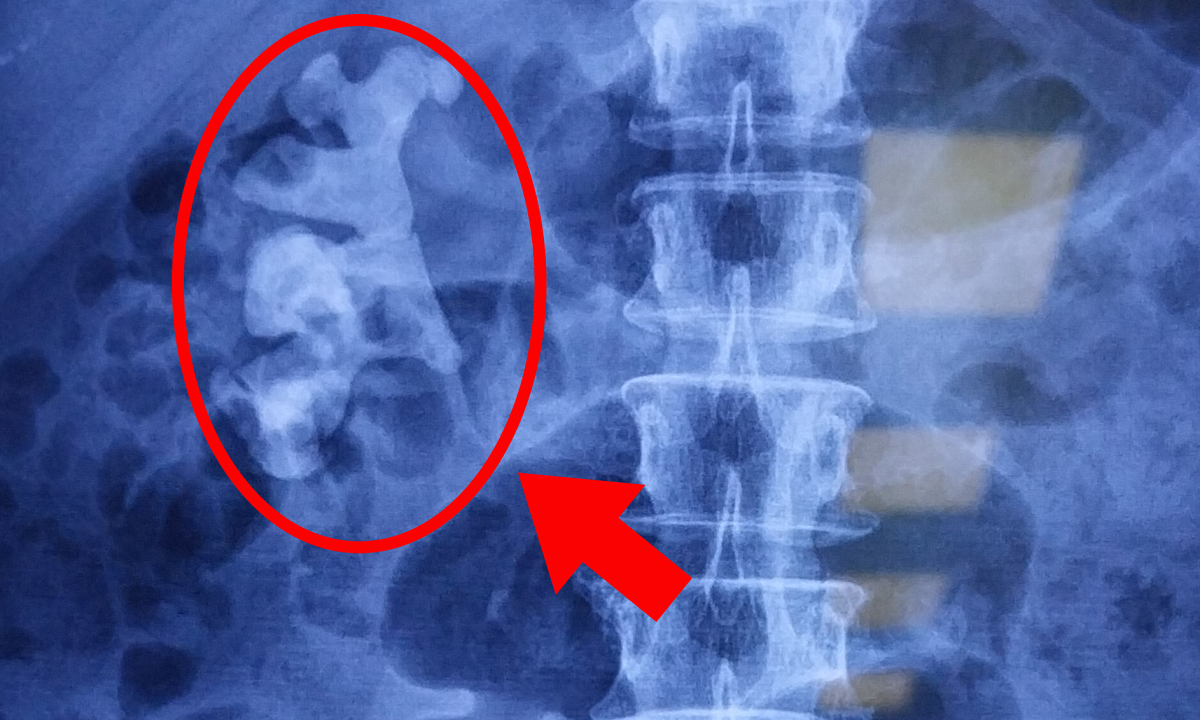
Ho Chi Minh City Ms. Hien, 36 years old, had a routine health check-up and a gastrointestinal endoscopy doctor discovered a submucosal tumor in her rectum.
On January 2, Dr. Pham Huu Tung, Deputy Director of the Center for Endoscopy and Endoscopic Surgery of the Digestive System, said that a small tumor measuring 0.9x0.7x0.5 cm was found under the patient's rectal mucosa and was indicated for removal to prevent possible complications.
The doctor used the Endoscopic Submucosal Dissection (ESD) technique to remove the entire tumor in about 30 minutes. The tumor was sent for testing to determine whether it was benign or malignant. The patient was discharged the same day and received home health care according to the doctor's instructions.
The biopsy results showed a benign tumor, a grade one neuroendocrine tumor. Dr. Tung said that this type of tumor originates from specialized cells of the body's neuroendocrine system. Due to some factors, the neuroendocrine cells change and no longer develop normally, forming a tumor. In the early stages, the disease has no symptoms and is often discovered by chance during an endoscopy for a health check.
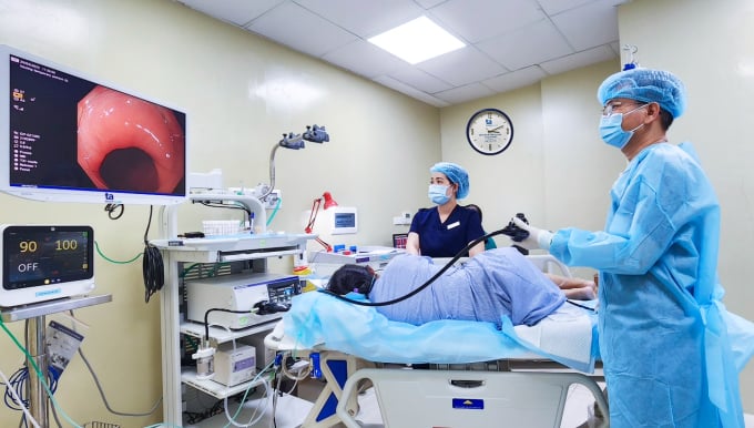
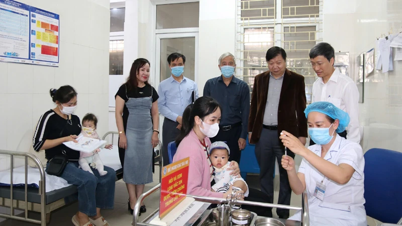
Doctor Huu Tung (blue shirt) performed a gastric endoscopy on a patient in October. Illustration photo: Provided by the hospital
Dr. Tung assessed that complete resection of the lesion is the optimal, minimally invasive method to help treat diseases of the gastrointestinal mucosa, precancerous lesions and early cancer in the stomach and colon. The method is performed entirely endoscopically, helping to limit complications such as perforation and bleeding.
Submucosal tumors are lesions that protrude into the lumen of the digestive tract (the lining of the digestive tract), and can occur anywhere from the esophagus to the rectum. This type of tumor is divided into many types such as stromal tumors, smooth muscle tumors, lipomas, granulosa cell tumors, neuroendocrine tumors, and lymphomas. Most tumors are benign, accounting for 85%.
"If submucosal tumors of the digestive tract are detected early, treatment is simple, economical, and highly effective," said Dr. Tung, adding that depending on the nature, size, and location of the tumor, the appropriate treatment method is chosen.
Normally, submucosal tumors less than 2 cm in size can be removed endoscopically by submucosal dissection. Large tumors that cause complications and have a high risk of cancer must be treated with invasive methods such as endoscopic full-thickness resection of the digestive tract, tumor resection via tunnel endoscopy, combined endoscopy and endoscopic tumor resection surgery, laparoscopic surgery or open abdominal surgery...
If submucosal tumors are not detected promptly, they can easily lead to complications such as gastrointestinal obstruction due to the large size of the tumor, hindering the flow of food in the stomach and digestion of food. Submucosal tumors can also cause bleeding.
The worst case scenario is a malignant submucosal tumor with the risk of stomach cancer. The disease is detected at a late stage, seriously affecting health, difficult to treat, and not very effective.
Doctors recommend that everyone live a healthy life and proactively have regular gastroscopy and colonoscopy to screen for gastrointestinal diseases, prevent cancer and dangerous complications.
People in high-risk groups such as genetic factors, relatives with cancer, obesity, HP bacteria infection, previous gastric surgery, people over 45 years old... need to consult a Gastroenterologist for endoscopy time to screen for the disease.
Quyen Phan
| Readers ask questions about digestive diseases here for doctors to answer |
Source link
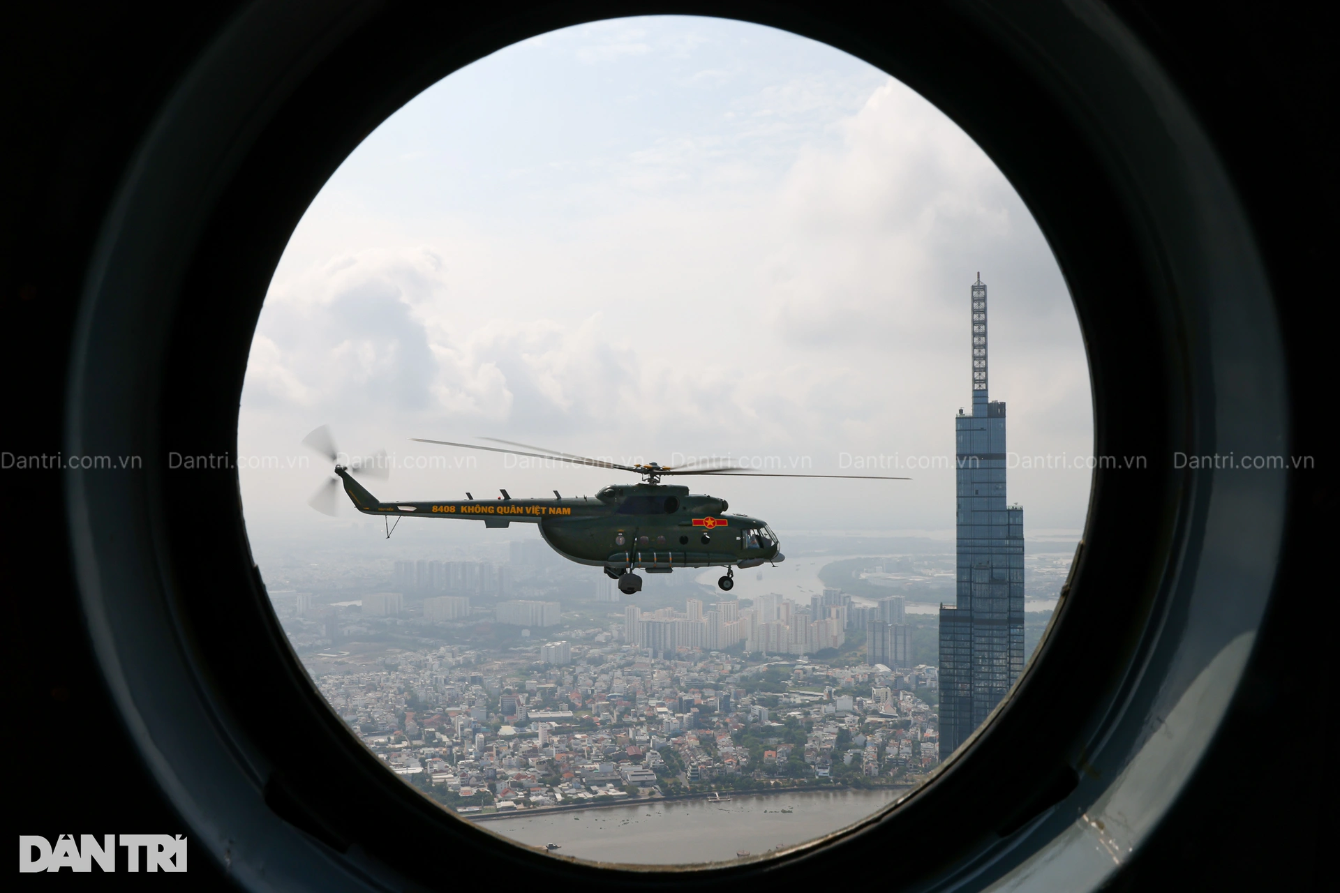
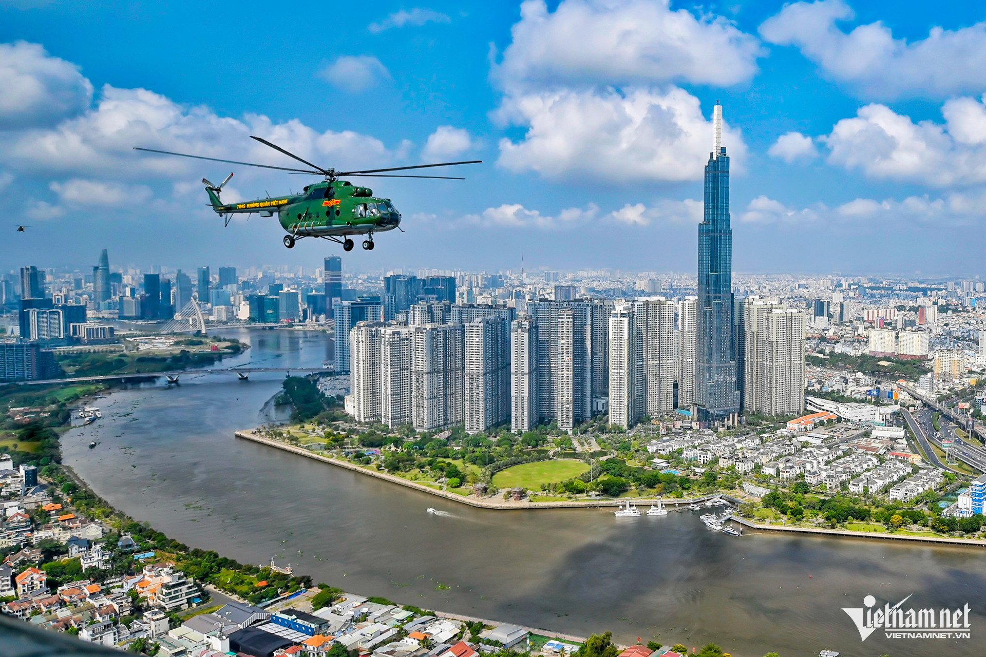
![[Photo] Helicopters and fighter jets practice in the sky of Ho Chi Minh City](https://vstatic.vietnam.vn/vietnam/resource/IMAGE/2025/3/28/3a610b9f4d464757995cac72c28aa9c6)
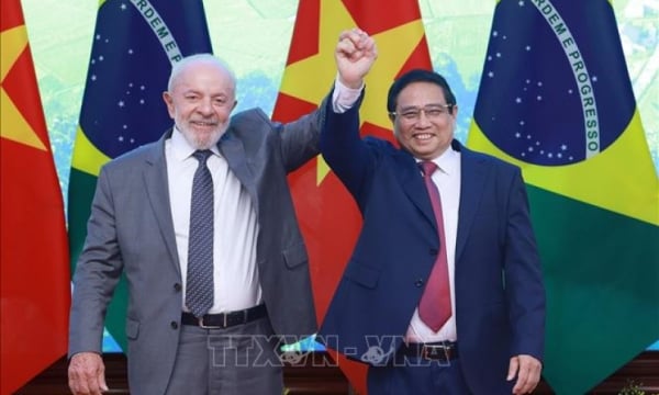
![[Photo] Prime Minister Pham Minh Chinh meets with Brazilian President Luiz Inacio Lula da Silva](https://vstatic.vietnam.vn/vietnam/resource/IMAGE/2025/3/28/41f753a7a79044e3aafdae226fbf213b)
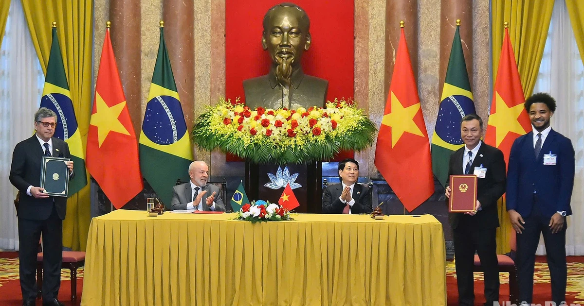
![[Photo] President Luong Cuong hosts state reception for Brazilian President Luiz Inacio Lula da Silva](https://vstatic.vietnam.vn/vietnam/resource/IMAGE/2025/3/28/56938fe1b6024f44ae5e4eb35a9ebbdb)
![[Photo] Flower cars and flower boats compete to show off their colors, celebrating the 50th anniversary of Da Nang Liberation Day](https://vstatic.vietnam.vn/vietnam/resource/IMAGE/2025/3/28/086d6ece3f244f019ca50bf7cd02753b)


Comment (0)