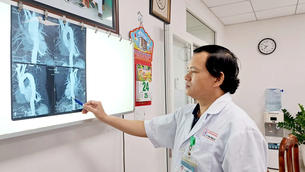
Patient NTK (57 years old, residing in Hoa Tho Dong ward, Cam Le district) accidentally discovered a lesion in the lower lobe of the left lung during a general health check-up. He had pneumonia 20 years ago and currently often has a dull pain in the left back.
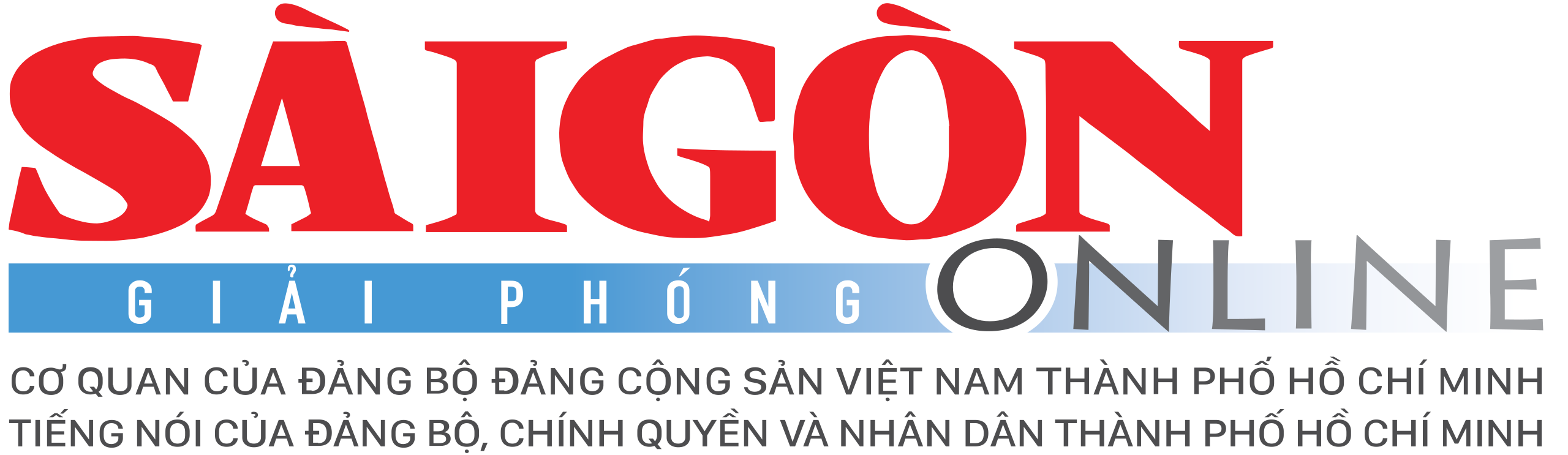
Chest CT scan with vascular reconstruction showed a 32x45mm lesion located in the lower lobe of the left lung. Notably, this mass was directly supplied by the thoracic aorta (diameter approximately 11mm) and drained venous blood into the left lower pulmonary vein. The patient was diagnosed with left intralobar sequestration.

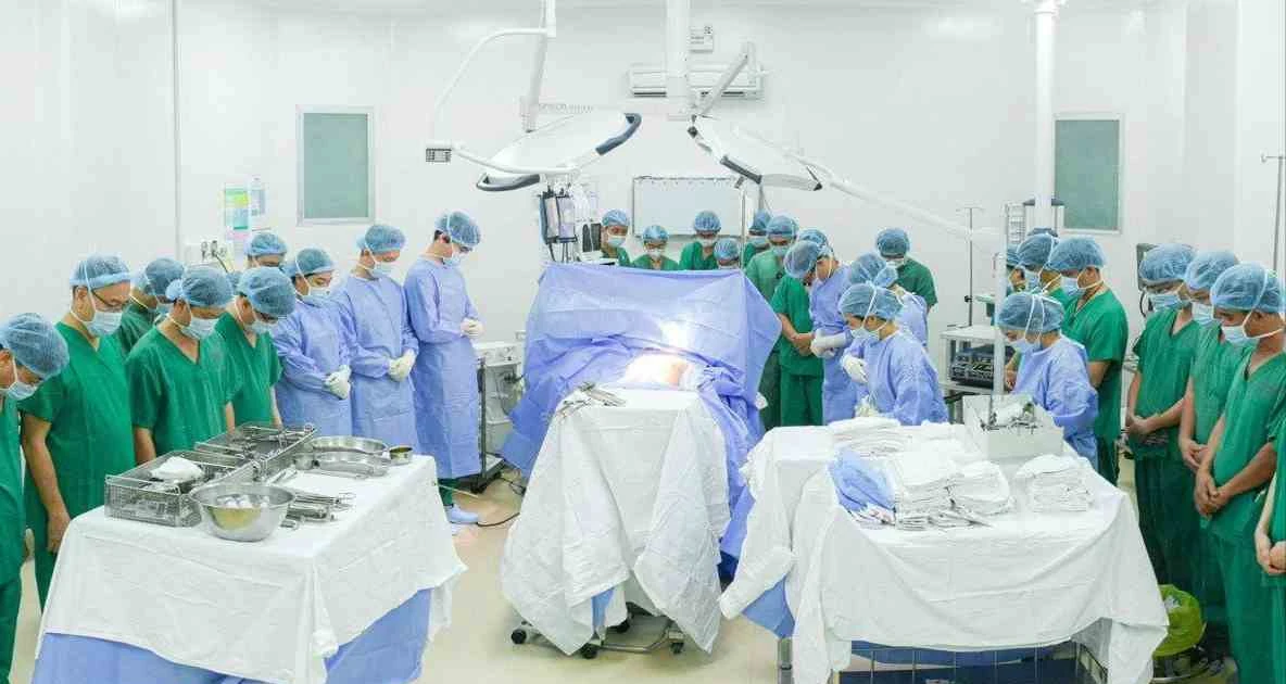
The doctors decided to perform a two-port thoracoscopic surgery to remove the isolated lung mass, in order to minimize chest wall trauma. After 2 hours of surgery, the team successfully removed the isolated lung mass and part of the parenchyma of the lower lobe of the left lung, while preserving as much of the remaining lung parenchyma as possible.
Dr. Than Trong Vu, Head of the Department of Thoracic Surgery, Da Nang Hospital (who directly performed the surgery) said that this case was quite challenging because the isolated pulmonary artery was large in size, while the patient was old, the artery wall was at risk of being brittle and prone to bleeding. Thanks to the meticulous coordination of the team, the surgery went smoothly. Currently, the patient is stable and is preparing to be discharged from the hospital.
According to Dr. Than Trong Vu, pulmonary sequestration is a rare congenital disease, accounting for 0.1% of congenital lung lesions. The disease is diagnosed in infancy. In adults, the diagnosis of pulmonary sequestration is rarely recorded. The progression of pulmonary sequestration can lead to recurrent pneumonia, lung abscess, cancer, severe bleeding that threatens the patient's life, heart failure due to increased cardiac output...
Source: https://www.sggp.org.vn/phau-thuat-noi-soi-cat-bo-phoi-biet-lap-hiem-gap-post790540.html
![[Photo] President Luong Cuong receives UN Deputy Secretary General Amina J.Mohammed](https://vstatic.vietnam.vn/vietnam/resource/IMAGE/2025/4/17/72781800ee294eeb8df59db53e80159f)
![[Photo] Warm meeting between the two First Ladies of the Prime Ministers of Vietnam and Ethiopia with visually impaired students of Nguyen Dinh Chieu School](https://vstatic.vietnam.vn/vietnam/resource/IMAGE/2025/4/17/b1a43ba73eb94fea89034e458154f7ae)
![[Photo] President Luong Cuong receives Kenyan Defense Minister Soipan Tuya](https://vstatic.vietnam.vn/vietnam/resource/IMAGE/2025/4/17/0e7a5185e8144d73af91e67e03567f41)
![[Photo] President Luong Cuong receives Lao Prime Minister Sonexay Siphandone](https://vstatic.vietnam.vn/vietnam/resource/IMAGE/2025/4/17/337e313bae4b4961890fdf834d3fcdd5)
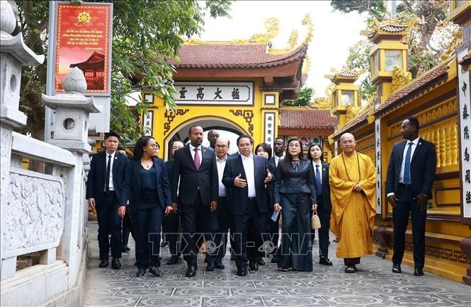
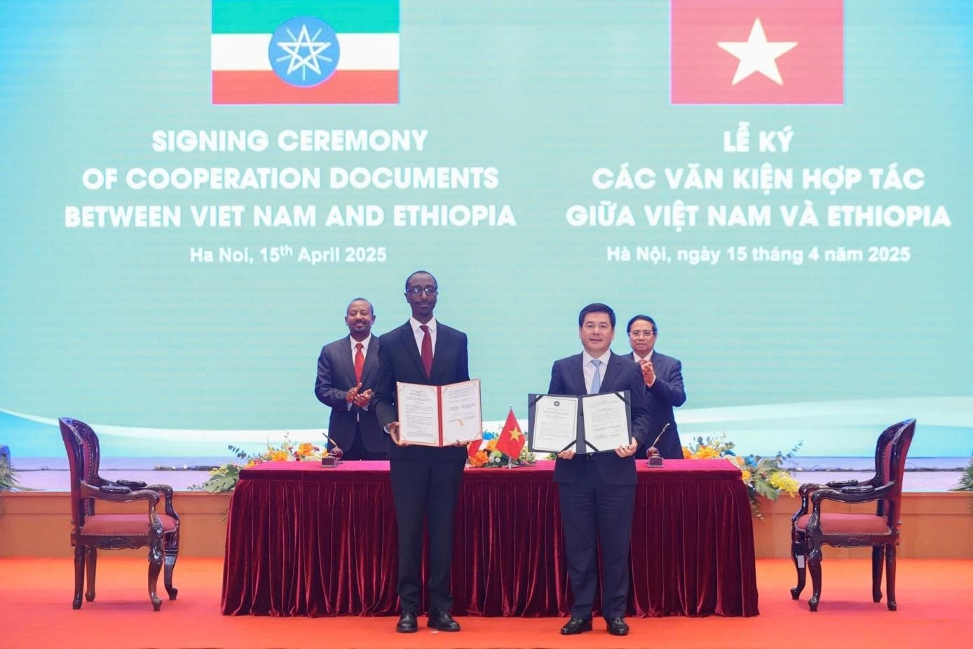
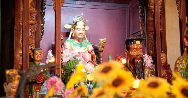
![[Photo] Prime Minister Pham Minh Chinh and Ethiopian Prime Minister visit Tran Quoc Pagoda](https://vstatic.vietnam.vn/vietnam/resource/IMAGE/2025/4/17/18ba6e1e73f94a618f5b5e9c1bd364a8)
![[Photo] Hundred-year-old pine trees – an attractive destination for tourists in Gia Lai](https://vstatic.vietnam.vn/vietnam/resource/IMAGE/2025/4/17/25a0b7b629294f3f89350e263863d6a3)


![[Video] Viettel officially puts into operation the largest submarine optical cable line in Vietnam](https://vstatic.vietnam.vn/vietnam/resource/IMAGE/2025/4/17/f19008c6010c4a538cc422cb791ca0a1)


Comment (0)