

Ho Chi Minh City – Ms. Thuy, 42 years old, was blind in her right eye and had only 2/10 vision in her left eye due to a tumor compressing the nerve; after surgery, her vision was restored.
Ms. Le Thi Thanh Thuy delayed brain tumor surgery for nearly three years due to caring for her young child and COVID-19. Since the beginning of 2023, her eyesight began to deteriorate significantly, and recently to the point of complete blindness, prompting her to seek treatment at Tam Anh General Hospital in Ho Chi Minh City.
On November 24th, Dr. Chu Tan Si, Head of the Neurosurgery Department at the Neuroscience Center, stated that the patient could only see with their left eye at a distance of 1-1.5 meters, and their vision was blurry. A 3 Tesla cranial MRI combined with deep tissue imaging (DTI) revealed a fairly large meningioma in the pituitary and suprasellar regions, approximately 5 cm in size. The tumor had grown to compress and completely envelop the optic nerve, causing blindness in the right eye and severely reduced vision in the left eye.
According to Dr. Tan Si, nearly three years after the tumor was discovered, the patient did not receive treatment, causing the tumor to grow and worsen. After a short time, the tumor continued to compress the convergence point of the two optic nerves, putting the patient at risk of blindness in both eyes.
The tumor also displaced the bundles of language and motor nerve fibers from their normal positions, invading both carotid arteries and both anterior cerebral arteries. These are important structures in the human brain.
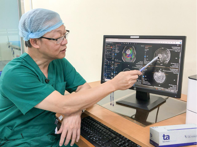
Doctor Tan Si assesses the approach to the tumor before surgery. Photo: Provided by the hospital .
The team decided to surgically remove the tumor with the assistance and guidance of the Modus V Synaptive AI-powered brain surgery robot.
To ensure the surgery was safe and precise, the surgical team created a 3D model of the brain structure using specialized robotic software. This allowed the doctors to clearly see the nerves, blood vessels, and other healthy structures within and around the tumor.
The surgeon performs a simulated surgery beforehand, proactively choosing a safe approach to the brain to remove the tumor without damaging surrounding nerve fibers and healthy brain tissue, thus preserving the patient's function after surgery.
The actual surgery followed the exact incision established during the simulation. The surgeon removed the tumor under the guidance and warning of abnormality indicated by robotic indicator lights. Most of the tumor was broken down and suctioned out using a Cusa ultrasound machine. A small portion of calcified tissue (due to the tumor being old) adhered to nerve structures, requiring manual removal by the surgeon.
After a 6-hour surgery, the tumor was completely removed, freeing both optic nerves and preserving the vascular structures within and around the tumor.
"The team didn't expect to recover the right eye because the patient had been blind for quite some time. However, a miracle happened; on the very day of the surgery, the patient's right eye could see faintly," said Dr. Tan Si.
Twenty-four hours after the surgery, the patient's vision improved significantly in both eyes. Upon checking the patient's vision, the doctor found that the left eye saw very clearly, and the right eye could see and correctly count the doctor's fingers.
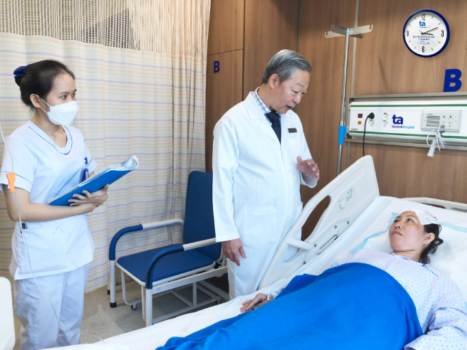
Doctor Tan Si examines Ms. Thuy after her successful surgery. Photo: Provided by the hospital.
Besides Ms. Thuy's case, Tam Anh Hospital has successfully performed surgery on nearly 100 cases of brain tumors and hemorrhagic strokes since deploying the AI Modus V Synaptive robot for brain surgery.
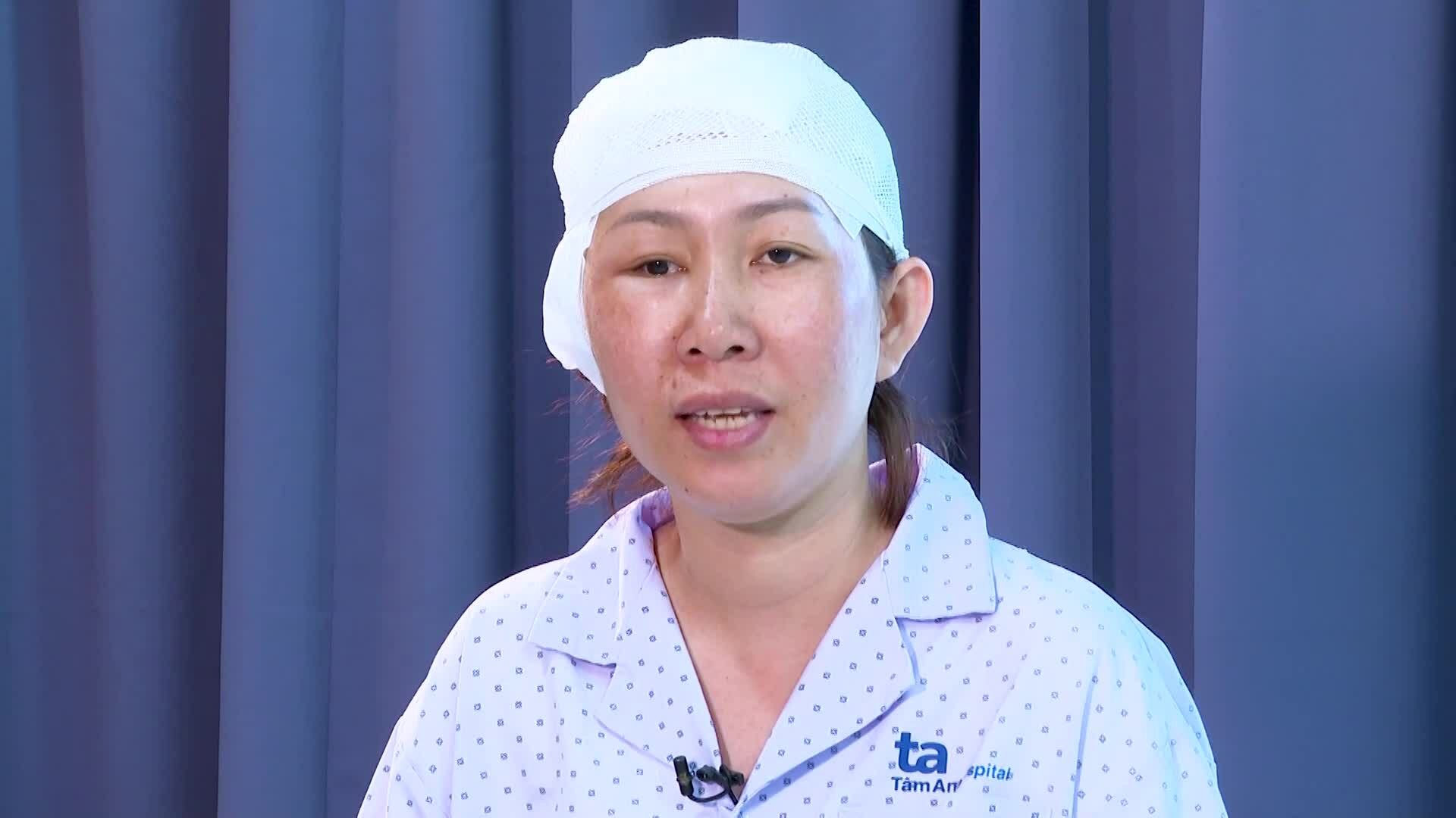
Doctor Tan Si and Ms. Thuy shared details about the surgery.
Peaceful
| Readers can post questions about neurological diseases here for doctors to answer. |
Source link
![[Photo] Prime Minister Pham Minh Chinh attends the Conference on the Implementation of Tasks for the Finance Sector in 2026](/_next/image?url=https%3A%2F%2Fvphoto.vietnam.vn%2Fthumb%2F1200x675%2Fvietnam%2Fresource%2FIMAGE%2F2026%2F01%2F06%2F1767699638245_ndo_br_dsc-0196-jpg.webp&w=3840&q=75)

![[Photo] Standing Committee member of the Party Central Committee Tran Cam Tu inspects preparations for the 14th Party Congress.](/_next/image?url=https%3A%2F%2Fvphoto.vietnam.vn%2Fthumb%2F1200x675%2Fvietnam%2Fresource%2FIMAGE%2F2026%2F01%2F06%2F1767695749033_image1122-1262-jpg.webp&w=3840&q=75)
![[Photo] Prime Minister Pham Minh Chinh receives Canadian Minister of International Development Randeep Sarai](/_next/image?url=https%3A%2F%2Fvphoto.vietnam.vn%2Fthumb%2F1200x675%2Fvietnam%2Fresource%2FIMAGE%2F2026%2F01%2F06%2F1767708661052_image123-3433-jpg.webp&w=3840&q=75)

![[Image] Fifth meeting of the Steering Committee for National Projects in the Railway Sector](/_next/image?url=https%3A%2F%2Fvphoto.vietnam.vn%2Fthumb%2F1200x675%2Fvietnam%2Fresource%2FIMAGE%2F2026%2F01%2F06%2F1767712857541_ndo_br_dsc-0581-jpg.webp&w=3840&q=75)


Comment (0)