
Ho Chi Minh City Ms. Anh, 29 years old, was diagnosed with metastatic brain cancer and treated in many places. Surprisingly, this time the brain surgeon discovered a benign tumor.
Two months ago, doctors at several hospitals diagnosed her with two large, malignant malignant tumors that were compressing her nervous system, predicting that the surgery would be risky, with the risk of blindness in both eyes and the inability to speak. Ms. Anh and her husband went to Tam Anh General Hospital in Ho Chi Minh City for examination, hoping that "there's still life, there's still hope".
On December 27, Dr. Huynh Tri Dung, Department of Neurosurgery, Center for Neuroscience , said that the patient came to the clinic in a state of confusion and anxiety, recently having symptoms of headache accompanied by a feeling of lightheadedness, difficulty speaking, and slow speech.
Magnetic resonance imaging (MRI 3 Tesla) shows two brain tumors located close to the ventricle wall, growing into the occipital horn and temporal horn of the left ventricle. The two tumors were approximately 3 cm and 4 cm in size, respectively. One tumor showed signs of internal hemorrhage, hemorrhage at the site where a biopsy had been taken previously at the hospital, and brain tissue edema.
Based on the nature, image of the lesion and the progression of the disease, Dr. Chu Tan Si, Head of the Department of Neurosurgery, initially assessed that the tumor could be benign. If the lesion is multifocal and malignant, it is not a high-grade malignancy.
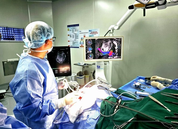
Doctors operated on Ms. Older brother. Photo: Provided by the hospital
With the above assessment, the team initially planned to perform a biopsy to assess the nature of the tumor and then perform chemotherapy and radiotherapy. At the same time, the team prepares a second plan: when performing brain surgery, directly approaching the tumor, the prognosis of the benign tumor is higher and can be easily removed, then the second tumor will be removed as well.
This assessment helps the surgeon choose a craniotomy that will allow both tumors to be removed. The patient undergo brain surgery using a robotic brain surgery system that combines a microsurgical microscope, neuro-navigation, and brain ultrasound.
Doctors combine MRI images of nerve fiber bundles on the robotic system, nerve positioning combined with neuro-ultrasound and microsurgery to accurately determine the location of the tumor before, during and after surgery. From there, they decide on the smallest skin incision and skull opening to access the tumor, minimizing damage to nerve fiber bundles and surrounding healthy brain tissue.
The incision was made in an arc shape 8 cm from the left temporal occipital. Under the microscope, the tumor was pinkish gray, tough, with many small hemorrhagic neovascular vessels. The surgeon dissected the tumor walls, used the Cusa machine to beat and aspirate the first tumor.
Under the guidance of the robot and neuronavigation, the doctors approached and removed the second tumor. Then, they placed a ventricular drainage tube outside and closed the skull cap.
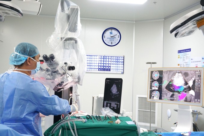
Ultrasound image checks the location of the injury after surgery. Photo: Provided by the hospital
After three hours of surgery, both tumors were completely removed. On the second day after surgery, Ms. He stood up and walked on her own. Three days after surgery, the biopsy results showed that the glioma (pilocytic astrocytoma) was benign.
"Holding the biopsy results, I felt like I was reborn a second time when I found out the tumor was benign," said Ms. Older brother.
Dr. Tan Si advises patients not to give up hope and stop treatment. In fact, treatment and pathology sometimes give different results than the initial diagnosis. Patients should follow the advice and treatment of specialized doctors.
Peaceful
* Patient's name has been changed
| Readers ask questions about neurological diseases here for doctors to answer |
Source link


Comment (0)