
Doctors at the Central Maternity Hospital have once again created a medical miracle, successfully saving a pregnant woman who had stopped circulating for more than 30 minutes and had to undergo four defibrillation shocks.
Medical news on February 15: Miracle of saving a pregnant woman's life at the Central Maternity Hospital
Doctors at the Central Maternity Hospital have once again created a medical miracle, successfully saving a pregnant woman who had stopped circulating for more than 30 minutes and had to undergo four defibrillation shocks.
This success is the result of the tireless efforts of a team of highly qualified doctors, quick response and synchronous coordination at a leading medical facility in the field of obstetrics.
Central Maternity Hospital creates miracle to save pregnant woman's life
The lucky woman who was saved was Ms. LTKN (34 years old, Long An ward, Thanh Hoa city, Thanh Hoa province). Previously, she was diagnosed with a third pregnancy, 38 weeks pregnant, with complications of central placenta previa, placenta accreta on the background of pregnancy-induced hypertension and a history of cesarean section. The surgery was performed when the woman was in critical condition.
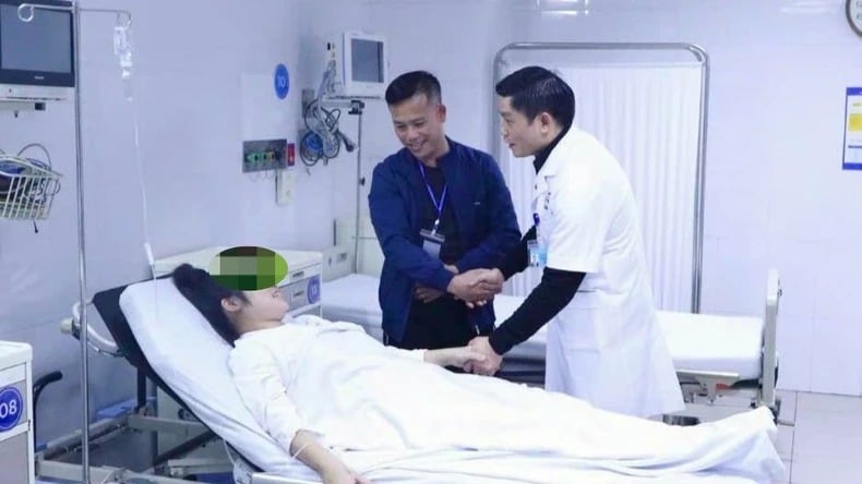 |
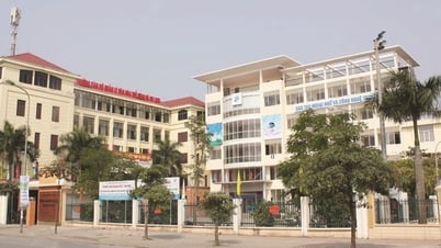
| Leaders of the Central Maternity Hospital visited and congratulated the mother. |
When visiting and encouraging the mother before discharge, Associate Professor, Dr. Nguyen Duy Anh, Director of the Central Maternity Hospital, sent his best wishes to the mother and baby.
He also expressed pride in the hospital's talented medical team, with more than 30 years of experience in the field of obstetrics. I understand that amniotic fluid embolism is a rare complication, but the mortality rate is very high, up to 85%, and often without warning signs.
On the morning of February 4, Ms. N. was taken to the operating room and the surgery initially went smoothly. The baby boy weighing 4.3 kg was born safely. However, immediately after the baby was born, the mother suddenly fell into critical condition due to amniotic fluid embolism.
Professor Bach Minh Thu, head of the Department of Surgery, Anesthesia and Resuscitation at the Central Maternity Hospital, said that when Ms. N's condition worsened, the mother suddenly turned purple, her blood pressure dropped rapidly, leading to circulatory arrest. Doctors diagnosed the patient with cardiac arrest and immediately took emergency measures.
"We cannot give up when there is still a chance to save the patient's life," Dr. Thu shared. The medical team activated the red alert for the entire hospital, mobilizing all the best human resources and experts to join the fight.
For more than 30 tense minutes, doctors performed continuous chest compressions, controlled ventilation with 100% oxygen, used cardiac drugs and emergency blood transfusion. After 20 minutes, the patient's heart rate began to return.
The battle to save the mother’s life lasted more than 2 hours, with the participation of the entire emergency team. After the heart beat again, the patient still had to face many problems such as blood clotting disorders, organ failure and concerns about brain damage due to prolonged cardiac arrest.
However, thanks to the tireless efforts of the doctors, the mother gradually regained consciousness and the problems were controlled. Master, Doctor Trinh Xuan Khanh - Pain Management Unit of the hospital said, this is truly a miracle when the mother recovered after a prolonged cardiac arrest.
Associate Professor, Dr. Nguyen Duy Anh emphasized that the success of this emergency case is not only a medical miracle but also a testament to the professionalism, courage and high responsibility of the medical team at the Central Maternity Hospital. This is the result of perfect coordination and high expertise of the entire medical team.
He also affirmed that the Central Obstetrics Hospital is always the place to receive and treat difficult obstetric cases, and is also the front line in obstetric examination, treatment and emergency care in Vietnam. "The success of this case is a clear demonstration of the remarkable development of the country's medicine," Prof. Anh said proudly.
Saving a patient with intestinal perforation due to red apple seeds
Recently, at Viet Duc Friendship Hospital, doctors successfully treated a patient with a perforated small intestine caused by a sharp foreign object. Patient NTK, 37 years old, from Thai Nguyen , was hospitalized with a generalized abdominal infection, also known as generalized peritonitis, due to swallowing a foreign object, a red apple seed.
Specialist Doctor II Le Nhat Huy, Deputy Director of the Center for Colorectal-Perineal Surgery, Viet Duc Friendship Hospital, said that the day before being admitted to the hospital, the patient had epigastric pain, then the pain gradually spread to the right iliac fossa. This symptom is very similar to acute appendicitis.
The patient was taken to a hospital in Thai Nguyen and had a CT scan, which showed suspected intestinal necrosis. The patient was immediately transferred to Viet Duc Friendship Hospital.
The patient was admitted to the hospital with a systemic infection and abdominal pain. After consultation, the doctors determined that this was a case of generalized peritonitis (infection of the entire abdominal cavity), and the patient was required to undergo emergency surgery.
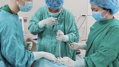
The patient underwent laparoscopic surgery, which showed cloudy fluid and pseudomembranes throughout the abdomen, no signs of inflammation in the appendix, and the small intestine loop at the junction of the small intestine and large intestine was inflamed and stuck together.
The patient’s entire abdomen was cleaned and drained. When performing an open surgery to examine the damage to the last part of the small intestine, the doctors discovered a hole in the intestinal wall, where a sharp foreign object was poking out. After removing the foreign object, it was determined to be a dried red apple seed – a familiar food to many people.
The patient was then treated for the perforated small intestine and the abdominal cavity was washed. After 8 hours, the patient regained consciousness, was transferred to the treatment room and recovered well.
According to the patient, Ms. K had previously eaten bird's nest stewed with red apples and accidentally swallowed the seeds. This is a very common situation, when many people do not pay attention when swallowing sharp foreign objects; because there is no choking or suffocation, the patient often subjectively thinks that the foreign object will be expelled.
Doctors warn that sharp foreign objects such as apple seeds, toothpicks, fish bones, chicken bones, etc. can cause damage to the digestive tract, especially the terminal ileum - where the small intestine connects to the large intestine. If not detected early, it can lead to intestinal perforation, peritonitis, severe infection, and even life-threatening.
In case of swallowing a sharp foreign object, the patient should not assume that the foreign object will be easily expelled. The patient should go to a medical facility within the first 4-6 hours to have a gastroscopy to remove the foreign object, avoiding complications of intestinal perforation.
Especially in the elderly or patients with poor health, abdominal infections can become more serious, easily leading to septic shock, poisoning and death.
To prevent this condition, people should practice the habit of eating slowly, chewing thoroughly, being especially careful when eating foods with hard seeds or bones, and not holding them in the mouth for too long. If you have swallowed a foreign object, especially a sharp object, you should quickly go to a medical facility for timely treatment.
Esophageal varices, a serious complication of cirrhosis
Esophageal variceal rupture is one of the severe complications of cirrhosis, which can cause gastrointestinal bleeding and is the main cause of increased mortality in patients.
Recently, Medlatec General Hospital has successfully performed esophageal variceal ligation, treating complications for a patient with cirrhosis. This is a typical case showing the danger of esophageal varices and the importance of timely intervention.
Patient NVT (40 years old, Hung Yen) has had chronic hepatitis B for the past 10 years and has been treated at Medlatec General Hospital. Six months ago, he was diagnosed with cirrhosis. Recently, he began to have symptoms of fatigue and jaundice, and decided to visit Medlatec for examination.
At the hospital, the patient's liver function test results were elevated. Abdominal ultrasound showed grade I fatty liver degeneration, enlarged gallbladder, and enlarged spleen. Esophagogastroduodenoscopy revealed grade II esophageal varices in the lower third of the esophagus, without red marks, and grade A gastroesophageal reflux, all of which are manifestations of portal hypertension.
Realizing the dangerous situation, doctors at Medlatec General Hospital assessed the risk of esophageal variceal rupture and gastrointestinal bleeding, which could threaten the patient's life.
Immediately, the doctors performed an endoscopic rubber band ligation of the esophageal varices. After the intervention, the patient was stable, ate well, and was prescribed outpatient treatment and discharged the same day.
In patients with cirrhosis, the stiffening of liver cells obstructs blood flow through the liver, increasing pressure in the portal veins and leading to changes in the flow of the portal venous system. As a result, the esophageal and gastric veins can become dilated. If they become too dilated, they can rupture and lead to severe bleeding.
MSc. Dr. Luu Tuan Thanh, Head of the Gastroenterology Department of Medlatec Healthcare System, said that patients with ruptured esophageal veins often have the following signs: vomiting a significant amount of blood, black stools, often feeling dizzy, or in severe cases, the patient may lose consciousness.
In addition, patients also have symptoms of cirrhosis such as jaundice, yellow eyes, easy bruising, bleeding. This is one of the main causes of gastrointestinal bleeding in patients with cirrhosis, and is a dangerous complication that threatens life.
When a patient experiences this condition, the doctor will prescribe emergency treatment and control the bleeding promptly. Once the bleeding has stabilized, the patient needs early intervention with esophageal variceal ligation to prevent dangerous complications.
Early endoscopic ligation of esophageal varices is an effective method to prevent the progression of varices and avoid vein rupture.
This method is indicated when the patient has symptoms such as vomiting blood, black stools, signs of blood loss due to bleeding such as low blood pressure, increased heart rate, decreased hemoglobin and red blood cell count. In addition, this technique is also applied to high-risk patients, such as large esophageal varices, red marks, or severe cirrhosis.
Esophageal variceal ligation is a simple technique and does not require complex equipment. It is performed quickly within 3-5 minutes. This method is highly effective, helping to prevent and treat gastrointestinal bleeding due to rupture or esophageal varices.
This method is indicated for severe patients. If not performed properly, it can cause serious complications such as bleeding or esophageal tearing.
Although esophageal variceal ligation is a simple technique, it requires the doctor to be highly skilled and perform the technique correctly. Therefore, doctors recommend that patients choose reputable medical facilities with experience in treating Gastroenterology to ensure safety and effectiveness of treatment.
Source: https://baodautu.vn/tin-moi-y-te-ngay-152-ky-tich-cuu-song-san-phu-cua-benh-vien-phu-san-trung-uong-d246577.html

![[Photo] Party and State leaders attend the special art program "You are Ho Chi Minh"](https://vphoto.vietnam.vn/thumb/1200x675/vietnam/resource/IMAGE/2025/5/18/6895913f94fd4c51aa4564ab14c3f250)
![[Photo] Many young people patiently lined up under the hot sun to receive a special supplement from Nhan Dan Newspaper.](https://vphoto.vietnam.vn/thumb/1200x675/vietnam/resource/IMAGE/2025/5/18/6f19d322f9364f0ebb6fbfe9377842d3)
![[Photo] Ready for the top competitions of Vietnamese table tennis](https://vphoto.vietnam.vn/thumb/1200x675/vietnam/resource/IMAGE/2025/5/18/9c547c497c5a4ade8f98c8e7d44f5a41)


Comment (0)