
Hanoi – Mr. Duong, 42 years old, experienced dull abdominal pain and fever. Doctors diagnosed him with a rare neuroendocrine tumor, with only about 150 cases recorded in world medical literature.
The results of the patient's endoscopic ultrasound and abdominal MRI showed a tumor protruding into the duodenum at the papilla of Vater (the final segment of the bile duct and pancreatic duct that empties into the duodenum). The tumor measured approximately 2x2 cm, with an ulcerated and bleeding surface.
On September 27th, Dr. Dao Tran Tien, Deputy Head of the Gastroenterology Department at Tam Anh General Hospital in Hanoi, diagnosed a carcinoid neuroendocrine tumor in the ampulla of Vater of the duodenum, which had not yet invaded the bile ducts or pancreas, requiring early intervention to avoid complications.
"Neuroendocrine tumors are mostly found in the gastrointestinal tract such as the stomach, small intestine, colon, rectum, appendix, and pancreas; they are rare in the ampulla of Vater," Dr. Tien said, adding that to date, only about 150 cases have been reported worldwide.
The incidence rate is approximately 0.3-1% of all neuroendocrine tumors in the gastrointestinal tract and less than 2% of all gastrointestinal cancers. In Vietnam, studies have noted that similar cases are quite rare.
According to Dr. Tien, the ampulla of Vater has a complex anatomical structure with many large blood vessels. If open surgery is performed on the duodenum, the patient is likely to experience complications such as pancreatic fistula, infection, scarring from the open incision, and a longer hospital stay and recovery time. Patients undergoing total pancreatectomy (removal of the entire duodenum and pancreas) may experience long-term health consequences and reduced quality of life. The team decided to perform endoscopic retrograde cholangiopancreatography (ERCP) to remove the ampulla of Vater tumor, preserving the digestive tract and reducing the risk of complications.
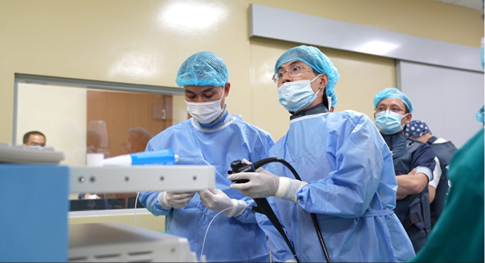
Doctor Tien (right) and his team perform endoscopic tumor removal on a patient. Photo: Provided by the hospital.
With the assistance of a C-Arm fluoroscopy system, the surgeon completely removed the tumor. The incision site was closed with specialized clips, promoting faster wound healing and preventing complications. The surgeon placed biliary and pancreatic stents to ensure bile duct recanalization and prevent complications such as edema, secondary bile duct obstruction, and acute pancreatitis.
Post-operatively, the patient's health stabilized, they were able to eat soft foods, and resumed normal activities. They were discharged two days later. The pathology results showed that the tumor was highly differentiated (i.e., low malignancy) and had been completely eradicated. The patient only needed follow-up and scheduled check-ups without requiring adjuvant treatment or chemotherapy.
Dr. Tien added that most tumors of the ampulla of Vater are malignant. If not detected and treated early, cancer cells can metastasize to other organs in the body, making treatment difficult. Furthermore, cancerous tissue can obstruct the ampulla of Vater, preventing bile and pancreatic juices from flowing into the small intestine for digestion, leading to bile duct obstruction, biliary tract infection, acute pancreatitis, biliary peritonitis, and death.
Neuroendocrine tumors are common in people aged 50-60, with women being more affected than men. The cause of the disease is currently unknown. Factors that increase the likelihood of developing the tumor include multiple endocrine neoplasia type one (MEN1), neurofibromatosis type one, and Von Hippel-Lindau syndrome (VHL).
Neuroendocrine tumors in the gastrointestinal tract progress silently, with no typical symptoms in the early stages. In later stages, clinical symptoms vary depending on the tumor location, easily leading to confusion with many other digestive diseases. The disease can only be completely cured when treated in the early stages.
According to Dr. Tien, primary tumors are usually small, so CT or MRI scans only have a sensitivity of 33% in diagnosis. To assess the detailed morphological characteristics of the tumor, doctors need the assistance of endoscopic ultrasound, which helps detect lesions deep beneath the thin layers of the digestive tract wall as well as surrounding lymph nodes and blood vessels. This allows for the assessment of regional lymph nodes and the extent of lesion invasion, enabling the development of an appropriate treatment plan.
Doctors advise that people experiencing unusual symptoms such as diarrhea, nausea and vomiting, abdominal pain, skin redness, and constipation should see a doctor for early diagnosis.
Trinh Mai
* Character names have been changed
| Readers can ask questions about digestive diseases here for doctors to answer. |
Source link


Comment (0)