
Thanks to ultrasound, doctors can screen and diagnose abnormalities early in pregnancy, thereby taking timely interventions to help babies be born safely and healthily.
In the field of obstetrics, ultrasound is considered a "third eye" that helps doctors access and see the baby inside the mother's womb.
Thanks to ultrasound, doctors can screen and diagnose abnormalities early in pregnancy, thereby taking timely interventions to help babies be born safely and healthily.
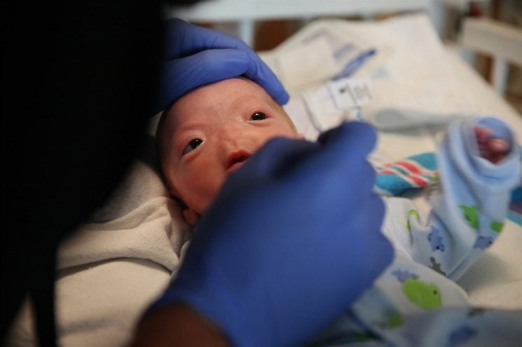 |
| Illustration photo. |
Therefore, experts recommend that pregnant mothers should pay attention and remember the important pregnancy check-ups and ultrasounds at 12, 22, 32 and 36 weeks.
According to the American Society for Reproductive Medicine (ASRM), the causes of recurrent miscarriages are uterine abnormalities, endocrine disorders, autoimmune diseases, genital tract infections, living environment, psychology, etc. Of which, about 60% of recurrent miscarriages are related to chromosomal abnormalities or genetic problems.
Accordingly, genetic testing before pregnancy can minimize the risks during pregnancy and increase the rate of giving birth to healthy babies. Depending on each type of genetic abnormality, the doctor will have appropriate treatment methods to help women get pregnant and give birth to healthy babies.
MSc. Ha To Nguyen, Director of the Center for Fetal Medicine, Tam Anh General Hospital, Ho Chi Minh City, said that ultrasound is an important method of screening pregnancy health in the field of maternity care.
Depending on the gestational age and stage, the doctor will prescribe appropriate ultrasound milestones for the pregnant mother. However, some important milestones that pregnant women need to pay attention to when going for a prenatal check-up for an ultrasound are 12, 22, 32 and 36 weeks.
The 12-week period helps detect high-risk pregnancies such as pre-eclampsia early, and is also a good time to screen for fetal abnormalities such as anencephaly, anencephaly, or severe complex abnormalities in the body such as external bowel, liver, or heart.
These abnormalities are very mild for the pregnant woman if terminated in the first trimester. Therefore, the 12-week period is extremely important in screening for early fetal abnormalities.
The 22-week ultrasound milestone is called the second trimester morphology ultrasound. About 50-60% of fetal abnormalities can be detected by ultrasound at this time.
Thanks to ultrasound, doctors can calculate gestational age, assess fetal diameter, assess amniotic fluid volume, weight and monitor fetal movement as well as comprehensively assess fetal structure including head, face, neck, chest, heart, skeletal system, abdomen, umbilical cord, and urinary system, thereby detecting important abnormalities in these organs.
Some abnormal cases will be treated by doctors at the Fetal Medicine Center using advanced techniques such as umbilical cord clamping in multiple pregnancies with complications, laser cautery in cases of twin blood transfusion causing one baby to have polyhydramnios, one baby to have oligohydramnios... In cases where the fetus has severe abnormalities, termination of pregnancy will be indicated.
The time when pregnant women will be assigned a 3D - 4D ultrasound to help detect abnormalities and support the diagnosis of fetal abnormalities. At this time, the mother can also see the baby's face on a 3D and 4D platform.
Ultrasound in the third trimester of pregnancy plays a very important role in re-examining fetal morphology to detect late-appearing abnormalities and plan for the baby at birth.
Not only that, this time also allows doctors to detect and evaluate fetal growth disorders including problems such as low birth weight, malnutrition, and intrauterine growth restriction.
There are two necessary times for ultrasound in the third trimester of pregnancy: 32 weeks and 36 weeks. In particular, ultrasound at the 36th week of pregnancy helps doctors detect growth restriction better than at the 32nd week.
In general, ultrasound milestones at weeks 12, 22, 32 and 36 are often applied to normal pregnancies. In case of abnormal pregnancies, the doctor will have specific instructions and perform additional ultrasound monitoring at other times.
According to Dr. Le Thanh Hung, Obstetrics and Gynecology Center, Tam Anh General Hospital, a high-risk pregnancy is a pregnancy that poses a risk to the mother or the baby or both, affecting the health of the mother and baby.
At the hospital, if the mother has diabetes or high blood pressure before pregnancy, the pregnancy will be monitored and sent to a cardiologist or endocrinologist to ensure health.
In addition, in cases where the pregnant mother is threatened with premature birth, the doctor will contact the Department of Neonatology for consultation so that the pregnant woman can be best prepared for the journey ahead. Or in cases where the fetus has abnormalities that can be treated after birth, obstetricians, cardiologists and related specialists will consult and provide the most optimal treatment regimen to prepare the baby for surgery after birth.
Fetal medicine is a medical specialty that aims to manage the health of mother and baby before, during, and after pregnancy. Fetal medicine has two client groups: mother and fetus.
Mother and fetus are two but one, for example, if the mother has a disease or health problem, it will be difficult to nurture a healthy fetus. On the contrary, a fetus with a disease needs intervention for chorionic villus sampling and amniocentesis, which must be done through the abdominal wall and the mother's uterus to be accessed.
Typical dangerous diseases such as congenital hemolytic anemia (Thalassemia) or couples with the disease gene will give birth to seriously ill babies, so their lives will be tied to the hospital for lifelong treatment.
With the development of modern medicine today, these dangerous diseases can be completely diagnosed before marriage. Therefore, examination before, during and after pregnancy is extremely important and necessary to help detect and control potential risks early, proactively protect pregnancy as well as plan for childbirth and save the baby's life.
Source: https://baodautu.vn/phat-hien-di-tat-som-bang-sieu-am-d220880.html
![[Photo] National conference to disseminate and implement Resolution No. 66-NQ/TW and Resolution No. 68-NQ/TW of the Politburo](https://vphoto.vietnam.vn/thumb/1200x675/vietnam/resource/IMAGE/2025/5/18/adf666b9303a4213998b395b05234b6a)


![[Photo] General Secretary To Lam visits exhibition of achievements in private economic development](https://vphoto.vietnam.vn/thumb/1200x675/vietnam/resource/IMAGE/2025/5/18/1809dc545f214a86911fe2d2d0fde2e8)


Comment (0)