
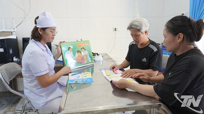
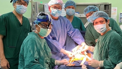
On November 29, speaking with Dan Tri reporter, specialist doctor Nguyen Trong Anh, Vice President of Ho Chi Minh City Sports Medicine Association, consultant of South Saigon Hospital, said that he and his colleagues had just performed surgery to save a woman who had suffered for 6 years from a rare disease.
Broken bones all over the body, 4 years of searching for the disease
The patient is Ms. LTL (34 years old, from Lam Dong ). Medical history shows that in 2017, about 2 months after giving birth to her second child, Ms. L. felt pain in her chest, ribs, and back, which then spread to her hips, right foot, and all over her body.
Despite being examined and treated everywhere, the patient did not get better but instead suffered more and more pain. There were times when the woman felt pain just breathing.

A rare disease called osteomalacia caused Ms. L. to suffer pain for 6 years (Photo: Hospital).
By 2019, Ms. L. was in a situation where she had difficulty walking, could not lift her legs, while she was mentally panicked and depressed because she did not know what disease she had. A hospital diagnosed her with metastatic cancer, and when she had an X-ray, she saw only broken bones everywhere, or suspected osteoporosis because of low bone density.
In early 2021, the patient met Dr. Ly Dai Luong, a lecturer at the Faculty of Medicine, Ho Chi Minh City National University, an endocrinologist. Based on the test results and symptoms, Dr. Luong discovered that the patient did not have osteoporosis but had osteomalacia.
This is a rare disease, in Vietnam only one case was reported in 2016, while the world has only recorded a few hundred cases. People with this disease will not see minerals and calcium in the bone structure, so the bones are very brittle and break easily.
Specific blood tests showed that the patient's blood phosphorus and calcium levels had dropped sharply. After careful examination, Dr. Luong determined that Ms. L's condition was caused by her kidneys.
Specifically, patients with tumors increase the secretion of FGF23 (a hormone that controls calcium-phosphorus metabolism), causing increased phosphorus excretion in the kidneys and reduced phosphorus and calcium absorption in the blood. To accurately locate the tumor, patients need to have a full-body PET-CT scan.

The patient was found to have a tumor in his heel that increased the secretion of FGF23, causing osteomalacia (Photo: Hospital).
"We consulted with Professor Chandran, a leading expert in metabolic bone disease in Singapore. She advised that the patient should have a PET-CT scan with the radioactive isotope Ga-68 (Dotatate gallium), but this substance is not available in Vietnam.
The patient had to go to Singapore for an X-ray in October, and finally discovered a tumor in the heel bone. The process of finding a place with a suitable endocrine radioactive isotope was very arduous, lasting for 2 years," said Dr. Trong Anh.
Enduring extreme pain while giving birth
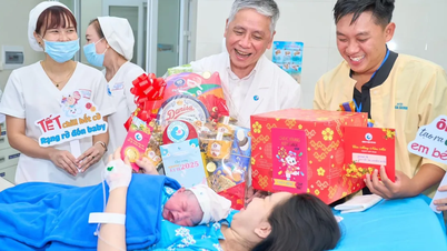
Dr. Ly Dai Luong shared that the time he met Ms. L. was in early 2021, when the patient had just given birth to her third child, even though she had known she had an unusual fracture 4 years before.
Talking to the doctor, the patient confided that she was in so much pain that she didn’t know when she would die, so she had to try to have more children. And after having 3 children, the patient was in so much pain that she couldn’t walk or even breastfeed.
"This may be the first patient in the world recorded to be pregnant and give birth while suffering from osteomalacia and multiple bone fractures in the body," said Dr. Luong.
For treatment, the patient was initially given phosphorus supplements, but had laxative side effects. During the 3 months of continuous diarrhea, while the patient was gradually deteriorating, a miracle happened, the pain improved well.

The patient was scheduled for surgery to carefully remove the tumor (Photo: Hospital).
Once the disease was "detected", the experts planned to surgically remove the tumor. Based on imaging, Ms. L's tumor was nearly 2.5cm long, occupying the entire horizontal diameter of the heel bone.
When the tumor was removed, the surgical site left a large bone cavity. The treatment team chose to use artificial bone to graft into the defect. The surgery lasted about 1 hour.
According to Dr. Trong Anh, the young mother’s tumor is most likely a benign mesenchymal tumor. But the sample will be sent to France to determine exactly what kind of tumor it is.

After removing the tumor, the bone defect in the patient's leg was grafted with artificial bone (Photo: Hospital).
In addition, it is uncertain whether the patient continues to produce FGF23 after tumor removal, so postoperative blood tests are needed to check.
Currently, Ms. L.'s health is gradually stabilizing. In the long term, the patient will be monitored for bone recovery and the patient's ability to absorb calcium and phosphorus (to see if osteomalacia recurs), as well as receive rehabilitation exercises.
Doctors recommend that rare diseases such as osteomalacia are not easy to detect. Therefore, patients with bone fractures and muscle weakness, in addition to seeing a musculoskeletal doctor, should go to an endocrinologist for examination, to determine the correct condition and treat it accurately and completely.
Source

![[Photo] President Luong Cuong receives President of the Senate of the Czech Republic Milos Vystrcil](/_next/image?url=https%3A%2F%2Fvphoto.vietnam.vn%2Fthumb%2F1200x675%2Fvietnam%2Fresource%2FIMAGE%2F2025%2F11%2F20%2F1763629737266_ndo_br_1-jpg.webp&w=3840&q=75)
![[Photo] Lam Dong: Panoramic view of Lien Khuong waterfall rolling like never before](/_next/image?url=https%3A%2F%2Fvphoto.vietnam.vn%2Fthumb%2F1200x675%2Fvietnam%2Fresource%2FIMAGE%2F2025%2F11%2F20%2F1763633331783_lk7-jpg.webp&w=3840&q=75)
![[Photo] National Assembly Chairman Tran Thanh Man holds talks with South Korean National Assembly Chairman Woo Won Shik](/_next/image?url=https%3A%2F%2Fvphoto.vietnam.vn%2Fthumb%2F1200x675%2Fvietnam%2Fresource%2FIMAGE%2F2025%2F11%2F20%2F1763629724919_hq-5175-jpg.webp&w=3840&q=75)


Comment (0)