[Excerpt from the entire article] Rare "egg" shaped extra-sinus renal pelvis stones were successfully treated by doctors of the Department of Nephrology and Urology - Thu Cuc International General Hospital with percutaneous nephrolithotomy using a mini tunnel, preserving maximum kidney function. Rare: "Egg" shaped stones in the kidney instead of the bladder With nearly 40 years of examining and treating diseases in the field of kidney and urology, Meritorious Doctor, Doctor CKII Pham Huy Huyen - Deputy Director of Thu Cuc International General Hospital, in charge of Nephrology and Urology, said: "It is rare to see a case of kidney stones protruding outside the sinus, round like a duck egg in a young patient." According to medical documents, large stones with round or oval shapes are often found in the bladder due to little compression and friction. Meanwhile, kidney stones are mainly irregular and rough in shape because they are limited by the narrow space of the kidney pelvis. However, the case of PKS (22 years old,
Hanoi ) surprised the urology experts at Thu Cuc TCI. The stone was diagnosed with a size of 3.0x4.1cm, occupying almost the entire space of the left kidney pelvis protruding outside the sinus, causing complications of renal pelvis dilation.
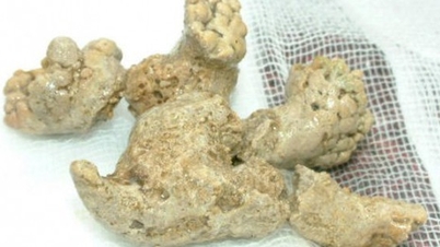
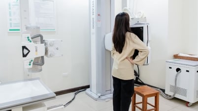
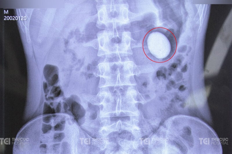 Diagnosis results of large stones in the renal pelvis outside the sinus (Photo: TCI)
 According to Dr. Huyen, the case of patient S. is not only special in shape but also in structure and age of onset, making the diagnosis and treatment process more complicated. In fact, at hospitals, kidney stones are common in middle-aged people and older, especially large stones. "However, S., at the age of 22, had a stone measuring up to 4.1 cm in the extra-sinus renal pelvis without causing pain or any obvious symptoms - this is very rare," Dr. Huyen affirmed. According to Dr. Huyen, the extra-sinus renal pelvis is a variation of normal anatomy. The extra-sinus renal pelvis is larger and more dilapidated than the intra-renal renal pelvis, which is surrounded by renal sinus fat. Based on research from the US National Library of Medicine of the US National Institutes
of Health , it is estimated that this variant can be found in about 10% of the population, while the majority of people have renal pelvis located entirely inside the kidney. "This shows more clearly that the 22-year-old patient with large, round stones located in the renal pelvis outside the sinus is a rare case," Dr. Pham Huy Huyen added. S.'s renal pelvis outside the sinus has a hole that flows into the ureter higher than normal and the ureteropelvic junction is narrow, causing the renal pelvis to become a "sink" to store urine. Most of the kidney sediment cannot be excreted, leading to the formation of large stones at a very young age.
Two lithotripsy sessions in 5 days with high-tech procedures. Faced with large stones and complex kidney malformations, the medical team considered many treatment options. Open surgery to remove stones with a 20-30cm incision on the skin is one of the options. However, this method can cause complications and long-term effects, especially for young patients at risk of stone recurrence due to malformed kidney structure. Therefore, percutaneous small tunnel lithotripsy is a superior solution with a small 5mm puncture on the skin, preserving maximum kidney tissue.
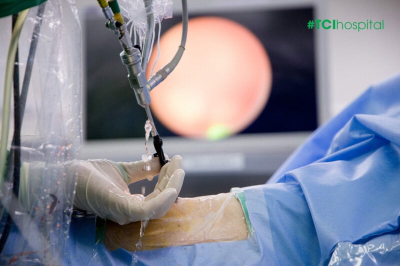 Percutaneous nephrolithotomy with minimal skin incision (Photo: TCI).
 This method uses high-power lasers and vacuum pressure to crush stones into small pieces and then suck them out, without the need for a large incision. However, due to the large size of the stones, it may be necessary to crush them 2-3 times. The medical team also calculated and developed an optimal protocol, using only one tunnel into the kidney for each crushing to remove the entire stone mass, reducing damage to the deformed kidney. Doctor Huyen shared: “The process of lithotripsy through the skin using a small tunnel is a big challenge, requiring high technique and strict adherence to standard time. On October 26, the first crushing was performed, removing half of the stone mass after more than 1 hour with about 23-25 liters of irrigation water. After 4 days, on October 30, the second crushing was performed to remove the remaining stones. By November 1, patient S. was walking normally, in stable health, expected to remove the drainage tube today and be discharged tomorrow.”
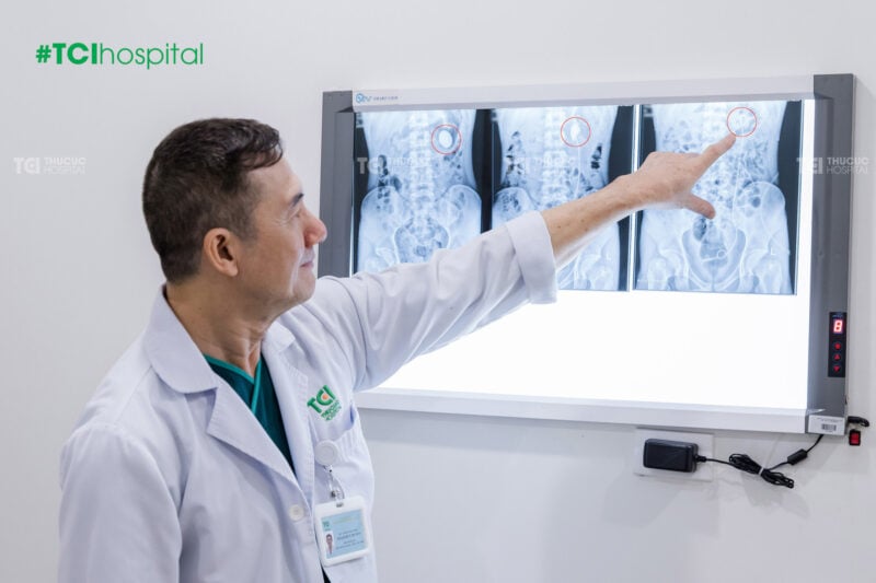 Results after 2 successful crushing sessions to remove large stones (Photo: TCI).
 PKS's case is a testament to the development of modern medicine in the treatment of kidney stones, especially in complicated cases such as renal pelvis malformations and large stones in young people. Thu Cuc International General Hospital has affirmed its pioneering role in applying cutting-edge treatment methods, bringing peace of mind and effective treatment options to patients.
 The patient's health is stable after percutaneous nephrolithotomy (Photo: TCI).
 With advanced techniques such as percutaneous nephrolithotomy with small tunnels, patients have new hope, not only for successful treatment but also for preserving kidney tissue and recovering health quickly.
Source: Dan Tri Newspaper Article link: https://dantri.com.vn/suc-khoe/hy-huu-soi-co-hinh-dang-nhu-qua-trung-xuat-hien-o-than-cua-chang-trai-22-tuoi-20241114220126817.htm
| Thu Cuc TCI International General Hospital masters advanced kidney and urinary stone lithotripsy technologies, prioritizing treatment with minimally invasive solutions, preserving kidney function for patients. Currently, customers have the opportunity to receive discounts of up to 30% on lithotripsy costs with all methods. Detailed information: https://benhvienthucuc.vn/tan-soi-cong-nghe-cao-danh-bay-soi-tiet-nieu/ Contact: 1900 55 88 92. |


![[Photo] Worshiping the Tuyet Son statue - a nearly 400-year-old treasure at Keo Pagoda](/_next/image?url=https%3A%2F%2Fvphoto.vietnam.vn%2Fthumb%2F1200x675%2Fvietnam%2Fresource%2FIMAGE%2F2025%2F12%2F02%2F1764679323086_ndo_br_tempimageomw0hi-4884-jpg.webp&w=3840&q=75)
![[Photo] Parade to celebrate the 50th anniversary of Laos' National Day](/_next/image?url=https%3A%2F%2Fvphoto.vietnam.vn%2Fthumb%2F1200x675%2Fvietnam%2Fresource%2FIMAGE%2F2025%2F12%2F02%2F1764691918289_ndo_br_0-jpg.webp&w=3840&q=75)


![[Infographic] 3 timelines of the "Quang Trung Campaign"](https://vphoto.vietnam.vn/thumb/402x226/vietnam/resource/IMAGE/2025/12/04/1764800248456_fb_ava-2.jpeg)


Comment (0)