
On March 23, Dr. Nguyen Ngoc Anh (Hepatology, Pancreatology Unit, Gia Dinh People's Hospital, Ho Chi Minh City) stated that after examination, doctors suspected a large tumor in the abdominal cavity and ordered blood tests and a multi-slice computed tomography (MSCT) scan of the abdomen for evaluation.
Clinical findings revealed a large tumor in the left lobe of the liver, occupying the entire abdominal cavity. The patient was diagnosed with hepatic hemangioma and admitted to the Hepatobiliary Pancreatic Unit for treatment.
Upon taking the patient's medical history, it was revealed that a tumor had been discovered in the abdomen during a medical examination eight years prior. Doctors recommended hospitalization for surgery to remove the tumor, but due to financial difficulties, the patient refused. Gradually, the tumor grew larger, compressing the inferior vena cava and forming collateral vessels in the anterior abdominal wall.
After admission, doctors at the Hepatobiliary and Pancreatic Unit collaborated with the hospital's DSA Unit to develop the best treatment plan for the patient. Although it was a benign tumor, its enormous size meant that open surgery would cause post-operative pain and cosmetic problems for the patient.
In addition, due to the long-term compression from the tumor, the patient had poor appetite and malnutrition, resulting in thin abdominal muscles that were prone to developing abdominal hernias later on. Ultimately, the doctors decided to perform laparoscopic surgery on the patient.

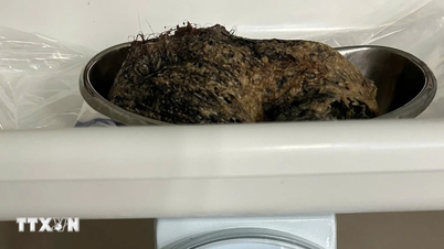
A tumor weighing over 5 kg was successfully removed from the patient's abdomen.
Prior to surgery, the DSA unit doctors assisted in embolizing the blood vessels supplying the tumor, aiming to reduce its size and minimize the risk of bleeding during the operation. Performing laparoscopic surgery required extreme caution because the tumor occupied almost the entire abdominal cavity, compressing other organs within the abdomen. Surgeons face numerous challenges when moving the liver, including the possibility of rupturing the tumor causing bleeding and the constant risk of damaging other organs in the abdominal cavity.
After careful consideration, the doctors safely removed the entire liver tumor, then made an open incision of about 20 cm above the pubic bone (corresponding to the cesarean section scar) to remove the entire tumor, weighing over 5 kg, from the abdomen. The surgery lasted approximately two hours. The patient's post-operative condition was stable, and she was discharged a few days later.
Dr. Ngoc Anh stated that hepatic hemangiomas are benign liver tumors, mostly small and asymptomatic. Patients only need regular check-ups to monitor the tumor's growth. In most cases, the tumor does not change in size or increases very little, only about 2 mm per year. Patients with hepatic hemangiomas should not worry too much; they just need to maintain a healthy lifestyle and a balanced diet to keep their liver healthy.
Source link


Comment (0)